
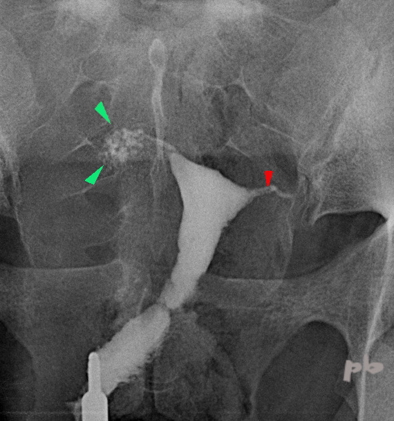
1- Endométriose tubaire – Hystérographie (HSG)
Multiples petites images d’addition en regard de la trompe droite (►). C’est l’image « en boule de gui », correspondant à une infiltration de la paroi tubaire +/- myométriale : salpingite isthmique noueuse dont l’endométriose est une des étiologies.
Trompe gauche (segment interstitiel ►).
1- Tubal endometriosis – Hysterosalpingography (HSG)
Multiple small additional images along the right fallopian tube (►) corresponding to infiltration of the tubal wall +/- myometrial wall : isthmic nodular salpingitis, of which endometriosis is one of the possible causes.
left fallopian tube (interstitial segment ►)
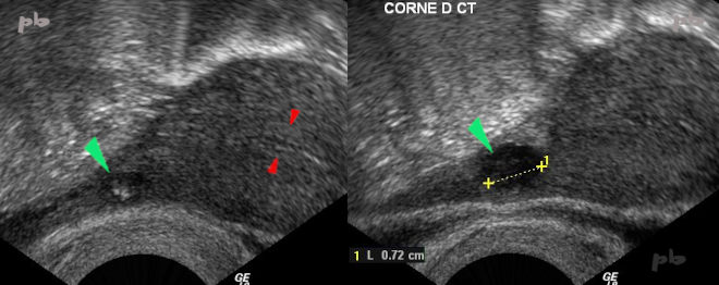
2- Endométriose tubaire – Echographie
Douleurs.
Un autre aspect en imagerie que la simple infiltration de la paroi tubaire.
Image hypoéchogène au niveau de la corne utérine droite (►) et du départ de la trompe. Ponctuations hyperéchogènes internes compatibles avec des dépôts d’hémosidérine.
Endomètre (►).
2- Tubal endometriosis – Ultrasound
Pain.
Another imaging aspect : hypoechoic image at the level of the right uterine horn (►) and the beginning of the fallopian tube.
Internal hyperechoic punctuations consistent with hemosiderin deposits.
Endometrium (►).
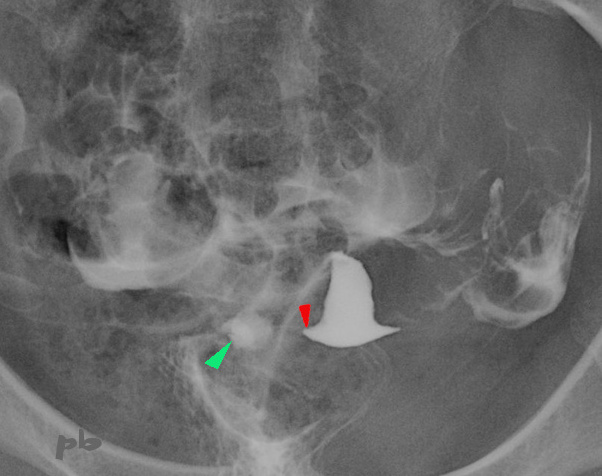
3- Endométriose tubaire – Hystérographie
(même patiente que 2)
L’image hypoéchogène de la corne utérine droite s’opacifie sur les clichés tardifs (►) formant une flaque de produit de contraste remplissant une cavité. Localisation sur le départ de la trompe (segments interstitiel +/- isthmique).
Corne utérine droite (►).
3- Tubal endometriosis – Hysterosalpingography
(same patient as 2)
The hypoechoic image of the right uterine horn becomes opaque on delayed images (►), forming a pool of contrast filling a cavity. Located at the beginning of the fallopian tube (interstitial +/- isthmic segments).
Right uterine horn (►).
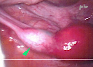
4- Endométriose tubaire – Coelioscopie
(même patiente que 2)
Vue coelioscopique.
Diagnostic confirmé à l’anapath.
4 – Laparoscopy
(same patient as 2)
Laparoscopic view.
Diagnosis confirmed by histopathology.

5- Endométriose tubaire – Echographie
Douleurs pelviennes cycliques de plus en plus intenses.
Image hypoéchogène d’allure hématique (►) située au niveau de la corne utérine droite, à proximité de la cavité endométriale (★).
Contours utérins (►).
5- Tubal endometriosis – Ultrasound
Increasingly intense cyclic pelvic pain.
Hypoechoic, hematoma-like image (►) located at the right uterine horn, near the endometrial cavity (★).
Uterine contours (►).
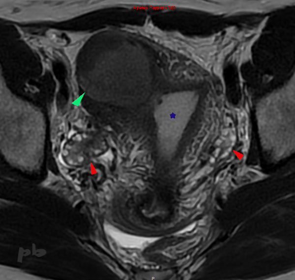
6- Endométriose tubaire – IRM
(même patiente que 5)
IRM coupe axiale T2.
L’image échographique apparait en signal intermédiaire en T2, hétérogène (►). Elle est bien intra-utérine, à proximité de la cavité endométriale (★) mais en-dehors de celle-ci.
Les ovaires (►) sont latéro-utérins, à distance de l’image.
6- Tubal endometriosis – MRI
(same patient as 5)
Axial T2-weighted MRI. The ultrasound image appears as an intermediate, heterogeneous signal on T2 (►). It is intrauterine, near the endometrial cavity (★) but outside of it. The ovaries (►) are lateral to the uterus, distant from the image.
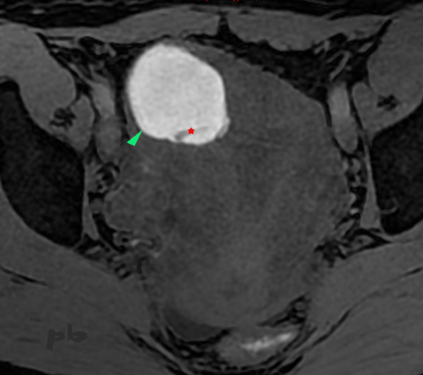
7- Endométriose tubaire – IRM
(même patiente que 5)
IRM coupe axiale T1 fatsat.
Image cornuale droite en hypersignal T1(►), confirmant sa nature hématique.
A noter une image linéaire à l’intérieur, en hyposignal : cloison de fibrine (★).
Une IRM réalisée 6 ans auparavant (non montrée) retrouvait exactement le même aspect.
7- Tubal endometriosis – MRI (same patient as 5)
Axial T1-weighted fat-saturated MRI.
Right cornual image with T1 hyperintensity (►), confirming its hemorrhagic nature. Note a linear hypointense image inside : a fibrin septum (★).
An MRI performed 6 years earlier (not shown) revealed exactly the same appearance.

8- Endométriose tubaire – Hystérosalpingographie (HSG)
(même patiente que 5)
Examen réalisé pour bilan d’infertilité.
Opacification à droite d’une cavité (►) correspondant à la trompe et à l’image échographique et IRM.
A noter que le diagnostic d’endométriose avait erré pendant plusieurs années, avec un épisode de diagnostic de grossesse cornuale à l’occasion d’une grossesse spontanée (qui s’était vite arrêtée).
8- Tubal endometriosis – Hysterosalpingography
(same patient as 5)
Examination performed for infertility assessment. Opacification on the right of a cavity (►) corresponding to the fallopian tube and the ultrasound/MRI findings.
Notably, the diagnosis of endometriosis had been missed for several years, including an episode where a cornual pregnancy was diagnosed during a spontaneous pregnancy (which quickly ended).
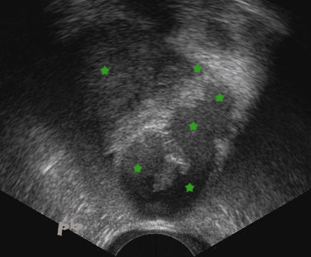
9- Endométriose tubaire – Echographie
Contexte d’endométriose sévère.
Volumineux hématosalpinx (★) à contenu liquidien échogène, hématique.
La GEU n’est pas la seule cause d’hématosalpinx.
9- Tubal endometriosis – Ultrasound
Context of severe endometriosis. Large hematosalpinx (★) with echogenic, hemorrhagic fluid content.
Ectopic pregnancy is not the only cause of hematosalpinx.
10-hématosalpinx – Echographie
Visualisation en temps réel du mouvement sanguin particulaire, isoéchogène(►), lors de la pression de la trompe par la sonde d’échographie.
10- Hematosalpinx – Ultrasound Real-time
Visualization of particulate blood movement, isoechoic (►), during pressure on the fallopian tube with the ultrasound probe.

11- Endométriose tubaire – Hématosalpinx
IRM coupe axiale T1 fatsat.
Trompe gauche (►) en hypersignal T1, hématique.
Il existait par ailleurs un nodule du torus (non montré).
11- Tubal endometriosis – Hematosalpinx
Axial T1-weighted fat-saturated MRI.
Left fallopian tube (►) with T1 hyperintensity, indicating hemorrhagic content. Additionally, there was a nodule on the torus (not shown).
12- Endométriose tubaire – Hématosalpinx – IRM
IRM séquence T2 dans le plan sagittal.
On suit sur les différentes coupes un volumineux hématosalpinx (★). Le liquide (sang) est de signal intermédiaire.
Ovaire homolatéral (►).
Col (►).
Vessie (★).
12- Tubal endometriosis – Hematosalpinx – MRI
T2-weighted MRI sequence in the sagittal plane.
A large hematosalpinx (★) is visible across the different slices. The fluid (blood) shows an intermediate signal.
Ipsilateral ovary (►).
Cervix (►).
Bladder (★).
© Dr Philippe BASSNAGEL – 2022