
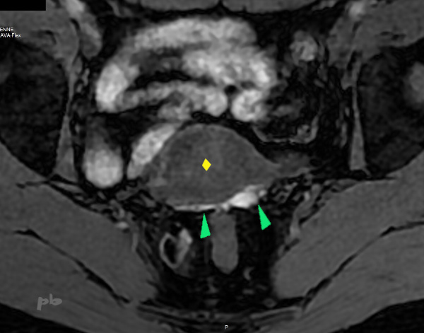
15 – Endométriose péritonéale postérieure
IRM coupe axiale T1 fatsat.
La face postérieure de l’utérus est tapissée par une bande en hypersignal T1, multinodulaire (►) : implants péritonéaux hémorragiques dans le cul-de-sac de Douglas.
Utérus (♦)
15 – Posterior peritoneal endometriosis
Axial T1-weighted fat-saturated MRI.
The posterior surface of the uterus is lined with a T1 hyperintense, multinodular band (►) : hemorrhagic peritoneal implants in the pouch of Douglas.
Uterus (♦)

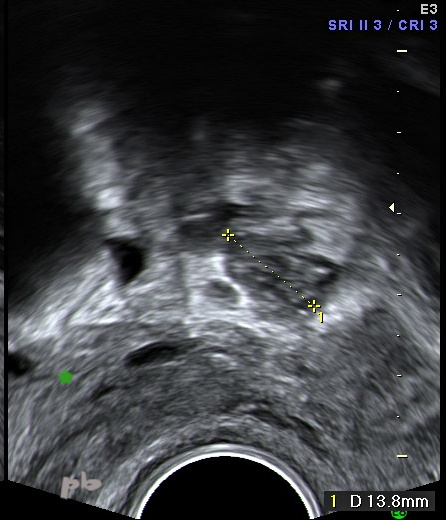
16 – Endométriose péritonéale antérieure
Echographie endovaginale.
Patiente atteinte d’une endométriose sévère.
-Image gauche : coupe sagittale.
Ponctuations hyperéchogènes (►) situées en regard du dôme vésical (► vessie vide).
-Image droite : coupe frontale du pelvis.
On retrouve les mêmes images au sein d’une plage hypoéchogène endométriosique (entre les croix), entre l’utérus (♦) et un kyste endométriosique ovarien gauche (★).
Ces ponctuations correspondent probablement à des plages hématiques.
En regard du dôme vésical, elles sont situées sur le péritoine du compartiment antérieur. Mais elles ne sont pas spécifiques d’implants endométriosiques, rencontrées par exemple aussi dans certaines infections (chlamydiose images 4, 6, 8).
16 – Anterior peritoneal endometriosis – Transvaginal ultrasound.
Patient with severe endometriosis.
–Left image : sagittal view. Hyper-echoic punctuations (►) located adjacent to the bladder dome (► empty bladder).
-Right image : frontal pelvic view. The same findings are seen within a hypo-echoic endometriotic area (between the crosses), between the uterus (♦) and a left ovarian endometriotic cyst (★).
These punctuations likely correspond to hemorrhagic areas. Adjacent to the bladder dome, they are located on the peritoneum of the anterior compartment. However, they are not specific to endometriotic implants, as similar findings can also be seen in certain infections (Chlamydiae images 4, 6, 8).

17 – Endométriose péritonéale antérieure
(même patiente que 16)
IRM coupe axiale T1 fatsat.
Hypersignaux antérieurs (►), en avant de l’utérus (♦), et situés en regard du dôme vésical, correspondant très probablement aux ponctuations hyperéchogènes décrites en échographie : implants hémorragiques péritonéaux endométriosiques.
Ces implants sont fréquemment rencontrés au cours de coelioscopie, souvent chez des patientes asymptomatiques. Ils sont par comparaison rarement visualisés en imagerie.
17 – Anterior peritoneal endometriosis
(Same patient as 16)
Axial T1-weighted fat-saturated MRI.
Anterior hyperintense signals (►), located anterior to the uterus (♦) and adjacent to the bladder dome, most likely corresponding to the hyper-echoic punctuations described on ultrasound : hemorrhagic peritoneal endometriotic implants. These implants are frequently encountered during laparoscopy, often in asymptomatic patients. In comparison, they are rarely visualized on imaging.
18 – Endométriose péritonéale
Patiente atteinte d’une endométriose sévère.
Échographie endovaginale.
Nodule (entre les croix) retrouvé en péri-sigmoidien, fixé.
Ovaire (★) à faible distance.
Nodule endométriosique péritonéal. Il a disparu après chirurgie sur les échographies ultérieures.
18 – peritoneal endometriosis
Patient with severe endometriosis. Transvaginal ultrasound.
Fixed nodule (between the crosses) found in the perisigmoid region. Ovary (★) located nearby. Peritoneal endometriotic nodule. It disappeared on subsequent ultrasounds after surgery.
© Dr Philippe BASSNAGEL – 2024