
Salpingites
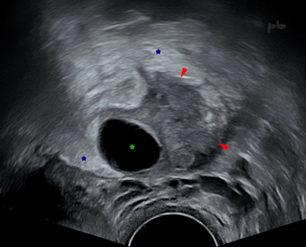
1 – Salpingite
Echographie endovaginale.
Pavillon tubaire épaissi (►). On voit les contours lobulés en rapport avec les franges tubaires, cernées par un peu de liquide.
Kyste paratubaire (★).
Hyperéchogénicité des tissus en périphérie (★), renforçant le contraste avec la trompe : inflammation.
1 – Salpingitis
Transvaginal ultrasound.
Thickened tubal infundibulum (►). Lobulated contours visible due to the tubal fimbriae, outlined by a small amount of fluid.
Paratubal cyst (★).
Hyper echogenicity of the peripheral tissues (★), enhancing contrast with the fallopian tube: inflammation.
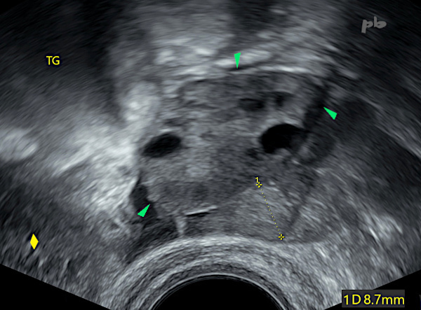
2 – Salpingite sur stérilet (DIU)
Echographie endovaginale.
La trompe (entre les croix) est épaissie (8,7 mm) et douloureuse à la palpation.
Ovaire gauche (►)
Utérus (♦)
2 – Salpingitis with IUD (Intrauterine Device)
Transvaginal ultrasound.
The fallopian tube (between the crosses) is thickened (8.7 mm) and painful on palpation.
Left ovary (►)
Uterus (♦)
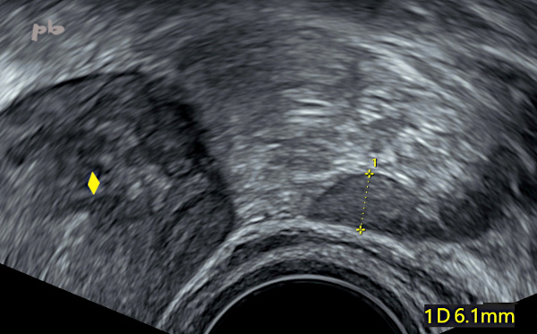
3 – Salpingite sur stérilet (DIU)
(Même patiente que 2)
Echographie endovaginale.
Contrôle après antibiothérapie.
La trompe (entre les croix) s’est normalisée (6,1 mm) et surtout les symptômes se sont améliorés.
Utérus (♦)
3 – Salpingitis due to IUD (intrauterine device)
(Same patient as case 2)
Transvaginal ultrasound.
Follow-up after antibiotic therapy.
The fallopian tube (between the crosses) has returned to normal (6.1 mm), and most importantly, the symptoms have improved.
Uterus (♦)
4 – Salpingite
Echographie endovaginale. Vidéo.
Douleurs pelviennes.
La trompe (►) est épaissie et douloureuse à la palpation. Au centre, muqueuse hyperéchogène épaissie et bien visible.
La trompe est suivie sur tout son trajet, depuis son émergence de l’utérus (♦) jusqu’au pavillon. Ce dernier est porteur d’un kyste (★) banal.
Follicule dominant sur l’ovaire gauche (étoile blanche).
Présence d’un épanchement (★) en postérieur, avec une cloison partielle que l’on voit flotter. A intégrer au tableau infectieux.
4 – Salpingitis
Transvaginal ultrasound. Video.
Pelvic pain.
The fallopian tube (►) is thickened and tender on palpation. In the center, there is a thickened, hyper-echogenic mucosa that is clearly visible.
The tube is traced along its entire path, from its emergence from the uterus (♦) to the fimbriae. The latter contains a common cyst (★).
Dominant follicle on the left ovary (white star).
Presence of a posterior effusion (★) with a partial septum that appears to be floating. This should be considered part of the infectious picture.
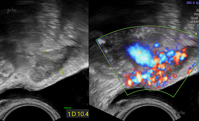
5 – Salpingite
Echographie endovaginale avec doppler.
Infection génitale haute après IMG (interruption médicale de grossesse).
La trompe (entre les croix) est épaissie (10,4 mm) et douloureuse à la palpation.
Hypervascularisation en doppler couleur.
Il existait un abcès tubo-ovarien de l’autre côté (non montré). Vessie en noir.
5 – Salpingitis
Transvaginal ultrasound with Doppler.
Upper genital infection following medical termination of pregnancy.
The fallopian tube (between the crosses) is thickened (10.4 mm) and painful on palpation.
Hypervascularization on color Doppler.
There was a tubo-ovarian abscess on the opposite side (not shown). The bladder is in black.
6 – Salpingite
(Même patiente que 5)
Echographie endovaginale.
Infection génitale haute après IMG (interruption médicale de grossesse).
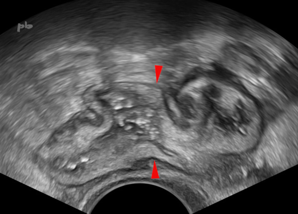
Le tableau infectieux comprenait un abcès tubo-ovarien gauche (non montré), une trompe droite épaissie, congestive (image précédente) et une inflammation pelvienne globale. On voit ici l’épaississement des parois sigmoïdiennes (►), surtout au niveau muqueuse et sous-muqueuse (la musculeuse hypoéchogène en périphérie a un aspect normal).
6 – Salpingitis
(Same patient as case 5)
Transvaginal ultrasound.
Upper genital infection following medical termination of pregnancy.
The infectious presentation included a left tubo-ovarian abscess (not shown), a thickened and congested right fallopian tube (previous image), and global pelvic inflammation. This image shows thickening of the sigmoid walls (►), particularly at the mucosal and submucosal levels (the hypoechoic muscularis layer on the periphery appears normal).
7 – Salpingite
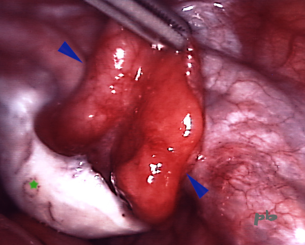
Coelioscopie.
Trompe congestive (►). Franges tubaires agglutinées.
Ovaire droit (★)
7 – Salpingitis
Laparoscopy.
Congested fallopian tube (►). Agglutinated fimbriae.
Right ovary (★)
8 – Salpingite sur stérilet (DIU)
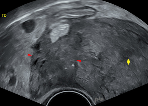
Echographie endovaginale. Coupe frontale.
DIU retiré la veille.
La trompe (►) est massivement épaissie et douloureuse à la palpation. Pas de plan de clivage visible avec l’utérus (♦) : adhérence.
Quelques petites images liquidiennes en périphérie en faveur de zones d’épanchement cloisonnées.
8 – Salpingitis due to IUD (intrauterine device)
Transvaginal ultrasound. Frontal view.
IUD removed the day before.
The fallopian tube (►) is massively thickened and painful on palpation. No visible cleavage plane with the uterus (♦): adhesion.
A few small fluid collections in the periphery, suggesting localized effusions.
9 – Salpingite
(Même patiente que 8)
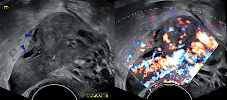
Echographie endovaginale. Coupe longitudinale
La trompe (entre les croix) est massivement épaissie (20,6 mm).
Quelques petites images liquidiennes en périphérie (►) en faveur de zones d’épanchement cloisonnées.
Importante hypervascularisation en doppler couleur.
9 – Salpingitis
(Same patient as case 8)
Transvaginal ultrasound. Longitudinal view.
The fallopian tube (between the crosses) is massively thickened (20.6 mm).
A few small fluid collections in the periphery (►), suggesting localized effusions.
Significant hypervascularization on color Doppler.
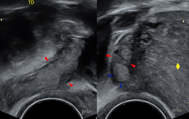
10 – Salpingite
(Même patiente que 8)
Echographie endovaginale. Coupe longitudinale à gauche et frontale à droite.
Traitement en cours.
La trompe (►) est mieux individualisée, moins épaisse.
Réapparition d’un plan de clivage avec l’utérus.
La muqueuse tubaire est anormalement visible, épaissie (►), car inflammatoire.
Utérus (♦)
10 – Salpingitis
(Same patient as 8)
Transvaginal ultrasound. Longitudinal view on the left and frontal view on the right.
Ongoing treatment.
The fallopian tube (►) is better defined and less thickened.
Reappearance of a cleavage plane with the uterus.
The tubal mucosa is abnormally visible and thickened (►) due to inflammation.
Uterus (♦)
© Dr Philippe BASSNAGEL – 2025