
Atlas d’images du pelvis féminin
Appareil urinaire
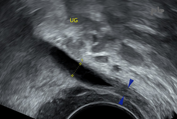
Des atteintes du tractus urinaire peuvent être rencontrées lors d’un examen pelvien réalisé à titre systématique ou dans un contexte symptomatique. L’échographie sera donc le 1er examen demandé le plus souvent, pour exploration du pelvis et/ou de l’appareil urinaire selon les cas.
Les localisations urétéro-vésicales de l’endométriose sont montrées dans le chapitre correspondant.
En cas de pathologie malformative gynécologique, la règle d’examiner les reins est bien connue. Mais l’inverse est vrai également (images 18 et 19).
Le cancer des voies urinaires est en-dehors des objectifs de cet atlas et ne sera pas abordé.
Toutes les techniques d’imagerie en coupe peuvent être utilisées. Cependant l’échographie a l’avantage de la simplicité et d’une grande fiabilité. Elle est à privilégier chez la femme, enceinte ou non.
Urinary tract conditions may be encountered during a pelvic examination performed routinely or in a symptomatic context. Ultrasound will therefore often be the first examination requested for the exploration of the pelvis and/or the urinary system, depending on the case.
The ureteral and bladder locations of endometriosis are shown in the corresponding chapter.
In cases of gynecological malformative pathology, the rule of examining the kidneys is well known. But the converse is also true (images 18 and 19).
Cancer of the urinary tract is beyond the scope of this atlas and will not be addressed.
All cross-sectional imaging techniques can be used. However, ultrasound has the advantage of simplicity and great reliability. It should be preferred in women, whether pregnant or not.
Docteur Philippe Bassnagel