

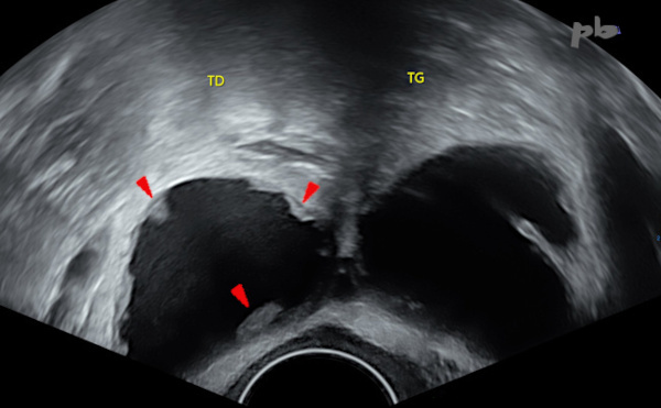
1 – Hydrosalpinx
Echographie endovaginale.
Hydrosalpinx typique : image tubulée, sinueuse, à contenu liquidien.
Absence de flux en mode B (pour les flux très lents) ou en doppler.
1 – Hydrosalpinx
Transvaginal ultrasound. Typical hydrosalpinx: a tubular, serpentine image with fluid content. No flow detected in B-mode (for very slow flows) or on Doppler.

2 – Vue coelioscopique
2 – Laparoscopic view
3 – Hydrosalpinx bilatéral
Echographie endovaginale.
Coupe transversale des 2 trompes.
Petites images pseudo-végétantes (![]() ) au sein de la lumière tubaire, de répartition régulière, donnant l’aspect classique de « roue dentée », caractéristiques de l’hydrosalpinx.
) au sein de la lumière tubaire, de répartition régulière, donnant l’aspect classique de « roue dentée », caractéristiques de l’hydrosalpinx.
Elles correspondent aux plis muqueux.
TD = trompe droite
TG = trompe gauche
3 – Bilateral Hydrosalpinx
Transvaginal ultrasound. Transverse section of both fallopian tubes. Small, pseudo-vegetative images ( ![]() ) within the tubal lumen, evenly distributed, creating the classic « gear wheel » appearance characteristic of hydrosalpinx. These correspond to mucosal folds.
) within the tubal lumen, evenly distributed, creating the classic « gear wheel » appearance characteristic of hydrosalpinx. These correspond to mucosal folds.
TD = Right fallopian tube
TG = Left fallopian tube
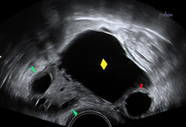
4 – Hydrosalpinx
Echographie endovaginale.
Kyste (♦) paraissant intra-ovarien (d’après l’angle de raccordement inférieur).
En-dessous sur le cliché, une image liquidienne avec l’aspect de « roue dentée » (![]() ), en faveur d’un hydrosalpinx.
), en faveur d’un hydrosalpinx.
Ovaire (![]() ).
).
4 – Hydrosalpinx
Transvaginal ultrasound.
A cyst (♦) appearing intra-ovarian (based on the lower attachment angle). Below in the image, a fluid-filled structure with a « gear wheel » appearance (![]() ), suggestive of hydrosalpinx.
), suggestive of hydrosalpinx.
Ovary (![]() )
)
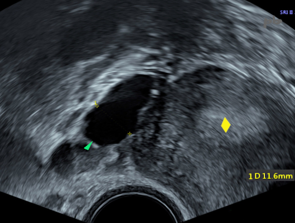
5 – Hydrosalpinx
(même patiente que 4)
Echographie endovaginale. Vidéo.
L’image de kyste ovarien (★) est en réalité en continuité avec l’autre structure liquidienne, ce que montre bien ce balayage échographique.
L’ovaire est distinct (★).
Il s’agit donc d’un hydrosalpinx, sans kyste ovarien associé. La portion la plus dilatée correspondant à la région ampullo-pavillonnaire.
5 – Hydrosalpinx (Same patient as image 4)
Transvaginal ultrasound. Video.
The apparent ovarian cyst (★) is actually continuous with the other fluid-filled structure, as demonstrated by this ultrasound sweep. The ovary is distinct (★). This is therefore a hydrosalpinx, with no associated ovarian cyst. The most dilated portion corresponds to the ampullary-infundibular region.
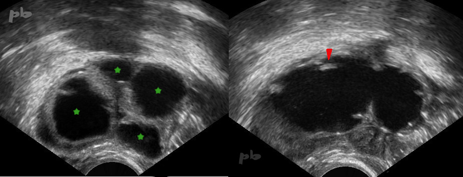
6 – Hydrosalpinx
Echographie endovaginale.
L’image de gauche montre plusieurs structures liquidiennes accolées les unes aux autres (★).
L’image à droite est réalisée après avoir tourné la sonde de 90 ° : les structures liquidiennes sont en continuité. On retrouve de plus des plis muqueux (►).
La trompe était pelotonnée sur elle-même, d’où cet aspect « multikystique ».
6 – Hydrosalpinx
Transvaginal ultrasound. The left image shows multiple adjacent fluid-filled structures (★). The right image was taken after rotating the probe by 90°: the fluid-filled structures are continuous. Additionally, mucosal folds are visible (►). The tube was coiled upon itself, resulting in this « multicystic » appearance.
7 – Hydrosalpinx
Echographie endovaginale.
Apparition d’un hydrosalpinx droit (►) lors d’une stimulation ovarienne.
Endomètre (♦)
7 – Hydrosalpinx
Transvaginal ultrasound. Appearance of a right hydrosalpinx (►) during ovarian stimulation.
Endometrium (♦)

8 – Hydrosalpinx
(même patiente que 7)
Echographie-doppler.
Apparition d’un hydrosalpinx droit avec une lumière tubaire irrégulière (►) lors d’une stimulation ovarienne. La paroi est épaissie (★), séquelle d’un processus inflammatoire.
Pas de flux enregistrable.
8 – Hydrosalpinx
(Same patient as case 7)
Ultrasound-Doppler. Appearance of a right hydrosalpinx with an irregular tubular lumen (►) during ovarian stimulation. The wall is thickened (★), a sequela of an inflammatory process. No detectable blood flow.
© Dr Philippe BASSNAGEL – 2023