
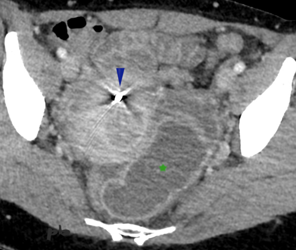
1 – Abcès tubo-ovarien
TDM coupe axiale.
Examen réalisé en urgence pour douleur et syndrome inflammatoire.
Présence d’un DIU en place (►).
En arrière et à gauche de l’utérus, image liquidienne tubulée (★) compatible avec un pyosalpinx dans le contexte de syndrome inflammatoire et DIU.
1 – Tubo-ovarian abscess
Axial CT scan.
Emergency examination performed for pain and inflammatory syndrome.
Presence of an IUD in place (►).
Behind and to the left of the uterus, a tubular fluid-filled image (★) consistent with a pyosalpinx in the context of inflammatory syndrome and IUD.
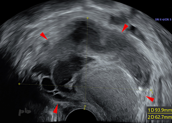
2 – Abcès tubo-ovarien
(même patiente que 1)
Echographie endovaginale réalisée dans les suites du scanner et après retrait du stérilet.
Masse complexe (►) associant des plages, d’aspect liquidien, échogènes, hypoéchogènes et anéchogènes. Ces dernières, à gauche sur l’image, font penser à des follicules et donc à la présence de l’ovaire.
Il est important de décomposer ce type d’image en éléments simples et reconnaissables : image suivante.
2 – Tubo-ovarian abscess
(Same patient as 1)
Transvaginal ultrasound performed following the CT scan and after IUD removal.
Complex mass (►) combining areas of liquid, echogenic, hypoechoic, and anechoic appearance. The latter, on the left side of the image, suggest follicles and thus the presence of the ovary.
It is important to break down this type of image into simple and recognizable elements: see next image.

3 – Abcès tubo-ovarien
(même patiente que 1)
Echographie endovaginale.
En modifiant le plan de coupe, il est possible de dérouler une structure tubulée à contenu liquidien échogène (partie déclive) et hypoéchogène : pyosalpinx (★).
D’autres images également à contenu liquidien hypoéchogène (►), ne sont pas reliées entre elles et correspondent à des plages abcédées.
Sous antibiothérapie, la patiente sera ponctionnée sous guidage échographique, avec une bonne évolution clinique.
3 – Tubo-ovarian abscess
(Same patient as 1)
Transvaginal ultrasound.
By adjusting the scanning plane, it is possible to unroll a tubular structure with echogenic (dependent part) and hypoechoic liquid content: pyosalpinx (★).
Other hypoechoic liquid-filled images (►) are not connected to each other and correspond to abscess areas.
Under antibiotic therapy, the patient will undergo ultrasound-guided drainage, with good clinical progression.
4 – Pyosalpinx
Echographie endovaginale – Boucle vidéo.
Balayage échographique dans le plan frontal sur l’annexe gauche.
Douleurs pelviennes depuis 48 heures, chez une patiente porteuse d’un DIU.
La trompe gauche (étoile blanche) est suivie sur tout son trajet, depuis le segment isthmique jusqu’au pavillon.
Son contenu est liquidien échogène (liquide épais). La paroi est un peu épaissie : 2 différences essentielles avec un simple hydrosalpinx.
Persistance de petites « végétations » correspondant aux plis tubaires.
L’ovaire gauche se situe en-dehors (►).
(La trompe droite était épaissie mais non dilatée.)
Utérus (♦)
4 – Pyosalpinx
Transvaginal ultrasound – Video loop.
Ultrasound sweep in the frontal plane over the left adnexa.
Pelvic pain for 48 hours in a patient with an IUD.
The left fallopian tube (white star) is followed along its entire course, from the isthmic segment to the infundibulum.
Its content is echogenic fluid (thick liquid). The wall is slightly thickened: two key differences from a simple hydrosalpinx.
Persistence of small « vegetations » corresponding to tubal folds.
The left ovary is located laterally (►).
The right fallopian tube was thickened but not dilated. Uterus (♦)
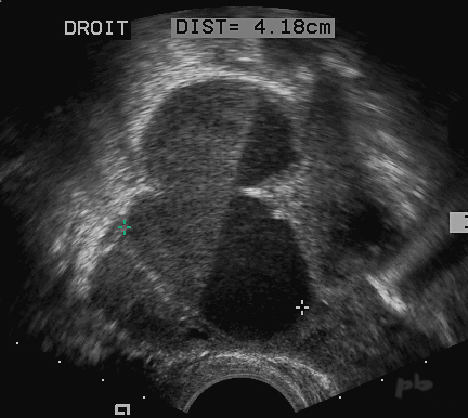
5 – Pyosalpinx
Echographie endovaginale.
Contexte de pelvipéritonite.
Trompe dilatée (croix), à contenu liquidien hypoéchogène, avec niveau liquide-liquide.
5 – Pyosalpinx
Transvaginal ultrasound.
Context of pelviperitonitis.
Dilated fallopian tube (crosses), with hypoechoic fluid content, and a fluid-fluid level.
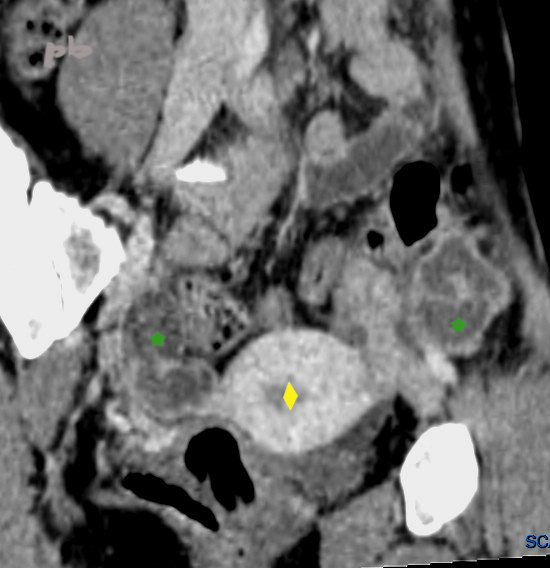
6 – Pyosalpinx bilatéral
TDM : reconstruction dans un plan frontal oblique.
Structure tubulée (★) de chaque côté de l’utérus (♦) : trompes dilatées à contenu liquidien et parois épaissies.
6 – Bilateral Pyosalpinx
CT scan: reconstruction in an oblique frontal plane.
Tubular structure (★) on each side of the uterus (♦): dilated fallopian tubes with fluid content and thickened walls.
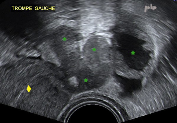
7 – Pyosalpinx bilatéral
(même patiente que 6)
Echographie endovaginale : coupe frontale à gauche.
Structure tubulée (★) à contenu liquidien isoéchogène à gauche de l’utérus (♦).
Encore plus à gauche, la portion distale de la trompe, à paroi épaisse et contenu liquidien anéchogène.
7 – Bilateral Pyosalpinx
(Same patient as 6)
Transvaginal ultrasound: frontal section on the left.
Tubular structure (★) with isoechoic fluid content to the left of the uterus (♦).
Further to the left, the distal portion of the fallopian tube, with thick walls and anechoic fluid content.
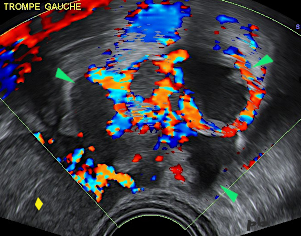
8 – Pyosalpinx bilatéral
(même patiente que 6)
Echographie endovaginale : coupe frontale à gauche en mode doppler.
Hypervascularisation des parois tubaires.
Pyosalpinx (►)
Utérus (♦)
8 – Bilateral Pyosalpinx
(Same patient as 6)
Transvaginal ultrasound: left frontal section in Doppler mode.
Hypervascularization of the tubal walls.
Pyosalpinx (►)
Uterus (♦)
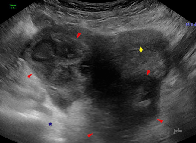
9 – Abcès tubo-ovarien
Echographie par voie sus-pubienne, coupe axiale.
Scanner réalisé ce jour pour un tableau de douleurs abdominales avec diarrhée. Contexte de carcinome mammaire. Complément échographique.
Volumineuse image pelvienne constituée par l’utérus (♦) et une masse (►) de contours floues, hétérogène. Hyperéchogénicité en arrière, en faveur d’un phénomène de renforcement postérieur (★) : masse liquidienne (liquide épais) ou tissu cellulaire « jeune » (tumoral) ?
9 – Tubo-Ovarian Abscess
Suprapubic ultrasound, axial section.
CT scan performed today for abdominal pain with diarrhea. Context of breast carcinoma. Complementary ultrasound.
Large pelvic image consisting of the uterus (♦) and a mass (►) with blurred contours, heterogeneous. Hyperechogenicity behind, suggesting a posterior reinforcement phenomenon (★): fluid mass (thick fluid) or tumor tissue?
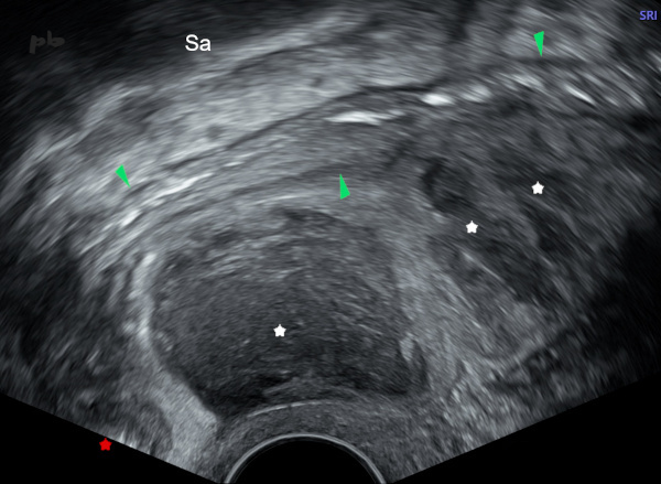
10 – Abcès tubo-ovarien
(même patiente que 9)
Echographie par voie endovaginale, coupe sagittale.
Anse sigmoïdienne (►) fixée au-dessus et en arrière à l’abcès (étoile blanche = plages liquidiennes purulentes ).
Pas d’épanchement dans le Douglas (★).
Sa = sacrum
10 – Tubo-Ovarian Abscess
(Same patient as 9)
Transvaginal ultrasound, sagittal section.
Sigmoid loop (►) fixed above and behind the abscess (white star = purulent fluid areas).
No effusion in the Douglas pouch (★).
Sa = sacrum
© Dr Philippe BASSNAGEL – 2025