
11 – Abcès tubo-ovarien
(même patiente que 9)
Echographie par voie endovaginale. Vidéo avec balayage dans le plan frontal pelvien droit.
Structure tubulée (étoile blanche) que l’on peut suivre sur la boucle vidéo, contenant un liquide échogène hétérogène de type pus. Au début de la séquence, présence d’images similaires mais sans continuité entre elles : plages abcédées cloisonnées.
Anse sigmoïdienne (►) fixée au-dessus à l’abcès.
L’utérus est à gauche (♦), sans plan net de séparation avec la masse (►). La mobilisation avec la sonde était également impossible (et douloureuse) : adhérence.
Enfin, il existe une importante hyperéchogénicité (★) en arrière de l’abcès et autour de l’anse digestive, trop importante pour un simple renforcement postérieur : inflammation des tissus cellulo-graisseux.
11 – Tubo-ovarian abscess (Same patient as case 9)
Transvaginal ultrasound. Video with sweeping in the right pelvic frontal plane.
A tubular structure (white star) traceable throughout the video loop, containing heterogeneous echogenic fluid consistent with pus. At the beginning of the sequence, similar images appear but without continuity between them: septated abscess areas.
Sigmoid loop (►) fixed above the abscess.
The uterus is on the left (♦), with no clear separation plane from the mass (►). Mobilization with the probe was also impossible (and painful): adhesion.
Finally, there is significant hyperechogenicity (★) behind the abscess and around the bowel loop, too pronounced for simple posterior acoustic enhancement: inflammation of the cellulofat tissues.

12 – Abcès tubo-ovariens bilatéraux
TDM après injection de produit de contraste.
Masses latéro-utérines à la fois solides et liquides (►).
En avant, la graisse est de densité augmentée et hétérogène (graisse « sale » ★).
Utérus (♦)
12 – Bilateral tubo-ovarian abscesses
CT scan after contrast injection.
Lateral uterine masses, both solid and liquid (►).
Anteriorly, the fat has increased and heterogeneous density (« dirty fat » ★).
Uterus (♦)
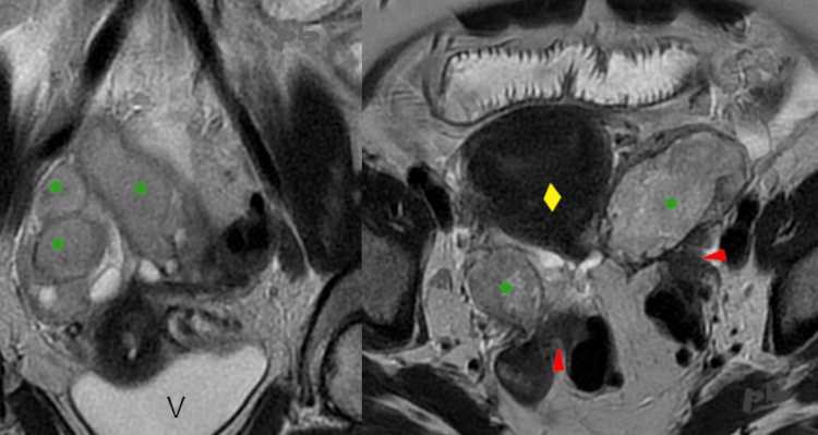
13 – Abcès tubo-ovarien
IRM coupes frontale à gauche et axiale à droite en pondération T2.
Pyosalpinx bilatéraux (★). Pus en signal intermédiaire.
Sur la coupe axiale, présence d’adhérences (►) avec le tube digestif.
Pas d’épanchement dans le Douglas, ce qui est habituel.
Utérus (♦)
V = vessie
13 – Tubo-ovarian abscess
MRI with frontal (left) and axial (right) T2-weighted slices.
Bilateral pyosalpinx (★). Pus with intermediate signal intensity.
On the axial slice, adhesions (►) are present with the digestive tract.
No fluid in the Douglas pouch, which is typical.
Uterus (♦)
V = bladder

14 – Abcès tubo-ovarien
Coelioscopie.
Volumineux pyosalpinx (►).
Utérus (étoile blanche)
Un peu de pus dans la cavité péritonéale (►).
Remerciements au Dr Patte
14 – Tubo-ovarian abscess
Laparoscopy.
Large pyosalpinx (►).
Uterus (white star).
A small amount of pus in the peritoneal cavity (►).
Acknowledgments to Dr. Patte
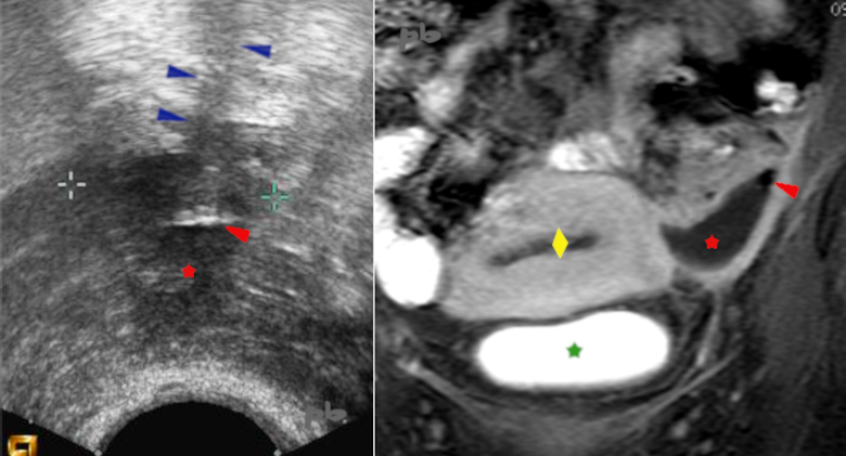
15 – Abcès post-opératoire
A gauche : échographie par voie endovaginale.
A droite : IRM coupe frontale après injection de gadolinium.
Présence d’une collection abcédée (★) du ligament large, apparue après césarienne.
On retrouve la présence de gaz au sein de la collection. En échographie, image linéaire hyperéchogène (►) avec artéfacts de répétition en arrière (►). En IRM, bulle sans aucun signal (►).
Vessie ★
Utérus ♦
15 – Postoperative abscess
Left: Transvaginal ultrasound.
Right: MRI frontal slice after gadolinium injection.
Presence of an abscessed collection (★) in the broad ligament, appearing after a cesarean section.
Gas is present within the collection. On ultrasound, a hyper-echogenic linear image (►) with posterior repetition artifacts (►). On MRI, a bubble with no signal (►).
Bladder ★
Uterus ♦

16 – Abcès pelvien
TDM avant (à gauche) et après drainage sous guidage scanographique (à droite).
Collection abcédée (★)
Drain (►)
16 – Pelvic abscess
CT scan before (left) and after CT-guided drainage (right).
Abscess collection (★)
Drain (►)
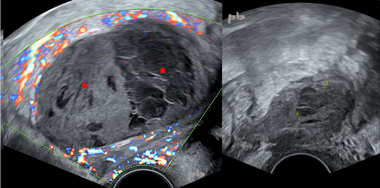
17 – Abcès pelvien
(même patiente que 16)
Echographie endovaginale avant (à gauche) et après drainage sous guidage scanographique (à droite).
Le contenu de l’abcès (★) apparait beaucoup plus hétérogène en échographie qu’en TDM. Absence de portions tissulaires comme le montre le doppler, éliminant une tumeur solide.
17 – Pelvic abscess (Same patient as case 16)
Transvaginal ultrasound before (left) and after CT-guided drainage (right).
The abscess content (★) appears much more heterogeneous on ultrasound than on CT. No tissue components are present, as shown by Doppler, ruling out a solid tumor.

18 – Endométriose et infection
A gauche : échographie par voie abdominale, coupe sagittale
A droite : IRM coupe axiale T1.
Patiente suivie pour infertilité, présentant une endométriose connue. Echographie 6 mois plus tôt retrouvant seulement des adhérence utéro-ovariennes gauches.
Aggravation des dysménorrhées.
L’échographie puis l’IRM réalisée quelques jours plus tard montrent un tableau d’endométriose sévère : volumineux kyste endométriosique ovarien gauche (★) en arrière de l’utérus (♦), un hydrosalpinx gauche et un nodule du torus (non montrés), et des adhérences.
Ve = vessie
18 – Endometriosis and infection
Left: Abdominal ultrasound, sagittal view.
Right: Axial T1-weighted MRI.
Patient monitored for infertility with known endometriosis. Ultrasound 6 months earlier only showed left utero-ovarian adhesions.
Worsening of dysmenorrhea.
The ultrasound, followed by an MRI performed a few days later, reveal severe endometriosis: a large left ovarian endometriotic cyst (★) behind the uterus (♦), a left hydrosalpinx, and a torus nodule (not shown), as well as adhesions.
Ve = bladder
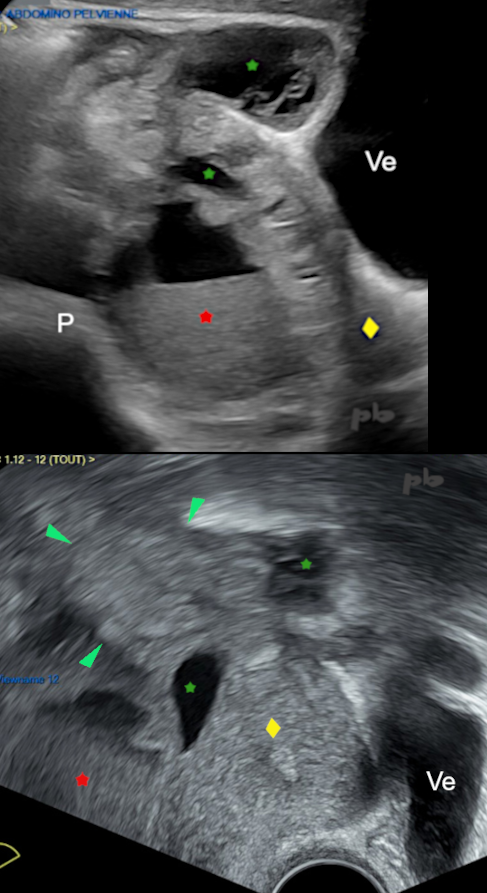
19 – Endométriose et infection
(même patiente que 18)
En haut : échographie par voie abdominale, coupe sagittale
En bas : échographie par voie vaginale, coupe sagittale
Bilan réalisé une semaine après le précédent (im18).
Douleurs de plus en plus intenses, et apparition d’un syndrome inflammatoire important. Sous antibiothérapie.
Le kyste endométriosique ovarien gauche (★) en arrière de l’utérus (♦) s’est modifié avec apparition d’un niveau liquide-liquide.
Les adhérences sont responsables de la formation de plusieurs collections (★) pelvienne à contenu liquidien épais. On repère aussi un paquet d’anses digestives (►) collées entre elles et aux structures de voisinage, associé à une hyperéchogénicité des tissus cellulo-graisseux : inflammation.
La patiente sera finalement opérée devant l’absence d’amélioration clinique sous antibiothérapie : faux kystes péritonéaux et inflammation. Les adhérences majeures empêcheront tout accès au pelvis.
-> Pelvi-péritonite sur endométriose.
Ve = vessie
P = promontoire
19 – Endometriosis and infection (Same patient as case 18)
Top: Abdominal ultrasound, sagittal view.
Bottom: Transvaginal ultrasound, sagittal view.
Assessment performed one week after the previous (image 18).
Increasingly intense pain and the appearance of a significant inflammatory syndrome. Under antibiotic therapy.
The left ovarian endometriotic cyst (★) behind the uterus (♦) has changed, with the appearance of a fluid-fluid level.
The adhesions are responsible for the formation of multiple pelvic collections (★) with thick liquid content. A cluster of bowel loops (►) is also identified, stuck together and to adjacent structures, associated with hyperechogenicity of the cellulofat tissues: inflammation.
The patient will ultimately undergo surgery due to the lack of clinical improvement under antibiotic therapy: peritoneal pseudocysts and inflammation. Major adhesions will prevent any access to the pelvis.
→ Pelvic-peritonitis due to endometriosis.
Ve = bladder
P = promontory
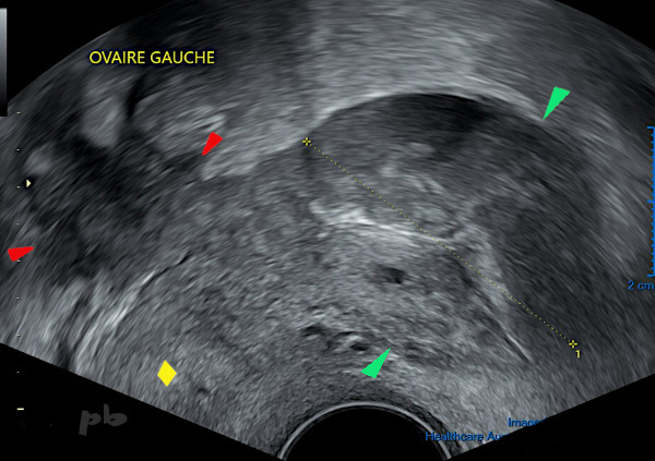
20 – Endométriose et infection
Patiente présentant des douleurs pelviennes sous pilule. Antécédents d’endométriose.
L’échographie endovaginale révèle un bloc utéro-ovarien, des kystes endométriosiques bilatéraux et un nodule endométriosique sur le torus (►), fixant en arrière le sigmoïde.
Kystes ovariens gauches d’allure endométriosique (►)
Ovaire gauche (croix)
Utérus (♦), sans interface visible avec l’ovaire : adhérences.
20 – Endometriosis and infection
Patient on the pill with pelvic pain. History of endometriosis.
The transvaginal ultrasound reveals a utero-ovarian mass, bilateral endometriotic cysts, and an endometriotic nodule on the torus (►), fixing the sigmoid colon posteriorly.
Left ovarian cysts with an endometriotic appearance (►)
Left ovary (crosses).
Uterus (♦), with no visible interface with the ovary: adhesions .
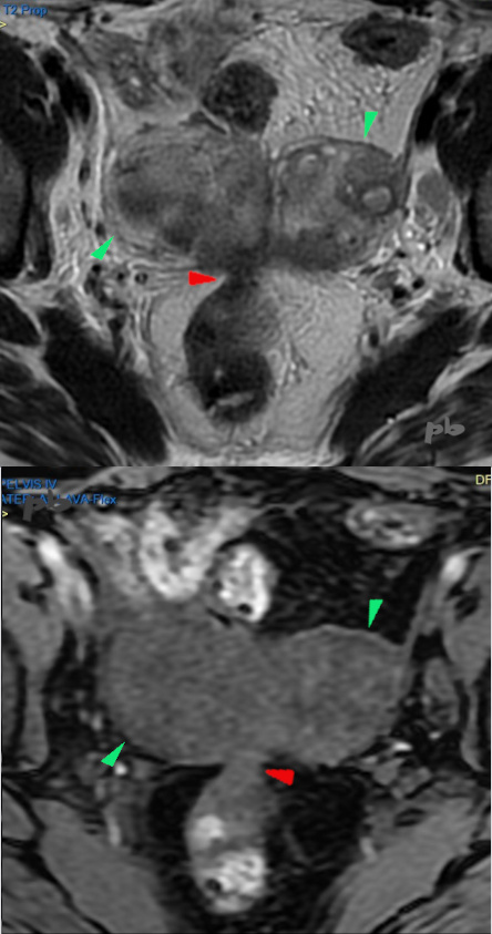
21 – Endométriose et infection
(même patiente que 20)
En haut : IRM coupe axiale T2
En bas : IRM coupe axiale T1
Bilan réalisé 3 jours plus tard (image 20).
Persistance des douleurs et syndrome inflammatoire.
On retrouve le nodule endométriosique sur le torus (►), fixant en arrière le sigmoïde.
En avant, 2 masses correspondant aux annexes. Le signal est hétérogène et relativement élévé en T2, et bas en T1. Ce qui avait été pris pour des kystes endométriosiques en échographie correspond en fait à 2 abcès tubo-ovariens.
Sous antibiothérapie, amélioration de la clinique et de la biologie.
Dans un contexte d’endométriose, une infection peut modifier un endométriome (image 19) ou faire confondre endométriome et abcès (image 21).
21 – Endometriosis and infection (Same patient as case 20)
Top: Axial T2-weighted MRI.
Bottom: Axial T1-weighted MRI.
Assessment performed 3 days later (image 20).
Persistent pain and inflammatory syndrome.
The endometriotic nodule on the torus (►) is visible, fixing the sigmoid colon posteriorly.
Anteriorly, there are 2 masses corresponding to the adnexa. The signal is heterogeneous and relatively high on T2, and low on T1. What was initially thought to be endometriotic cysts on ultrasound actually corresponds to 2 tubo-ovarian abscesses.
Under antibiotic therapy, clinical and biological improvement.
In the context of endometriosis, an infection can alter an endometrioma (image 19) or lead to confusion between endometrioma and abscess (image 21).
© Dr Philippe BASSNAGEL – 2025