

21 – Migration de DIU – ASP
Fils de DIU non vus cliniquement.
Echographie négative.
Réalisation d’un ASP retrouvant le DIU (►) au niveau de l’hypocondre gauche.
21 – IUD migration – Abdominal X-ray
IUD strings not clinically visible.
Negative ultrasound.
Performance of an abdominal X-ray showing the IUD (►) in the left hypochondrium.

22 – 2 DIU
ASP
Patiente ménopausée non traitée, porteuse d’un DIU au cuivre. Examen clinique anormal.
L’échographie retrouve un DIU cervical, avec branches latérales perforantes. Doute sur la présence d’un 2ème stérilet au niveau du fond cavitaire.
ASP réalisé dans les suites de l’écho, qui confirme l’existence de 2 DIU intra-pelviens.
22 – 2 IUDs
Abdominal X-ray
Postmenopausal patient, untreated, with a copper IUD. Abnormal clinical examination.
Ultrasound reveals a cervical IUD with perforating lateral arms. Suspicion of a second IUD in the uterine fundus.
An abdominal X-ray was performed following the ultrasound, confirming the presence of 2 intra-pelvic IUDs.
23 – DIU – Sac gestationnel
Echographie endo-vaginale – Coupe sagittale
Utérus rétrofléchi.
Présence d’un sac gestationnel (►) intra-utérin et d’un DIU abaissé (►), en position cervico-isthmique.
Fond utérin (♦).
23 – IUD – Gestational sac
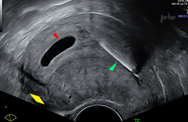
Transvaginal ultrasound – Sagittal view
Retroflexed uterus.
Presence of an intrauterine gestational sac (►) and a lowered IUD (►), in a cervico-isthmic position.
Uterine fundus (♦).
24 – Grossesse de 13 SA
Echographie sus-pubienne.
Visualisation d’un DIU (►) en coupe frontale jouxtant un sac gestationnel.
Pas de décollement.
Tête fœtale (►).
Liquide amniotique (♦).
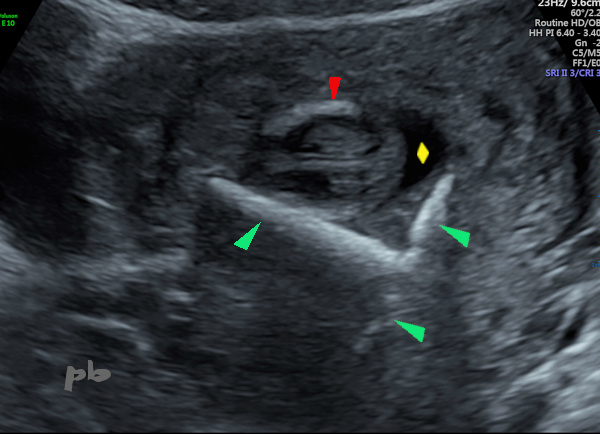
24 – 13-week pregnancy
Suprapubic ultrasound.
Visualization of an IUD (►) in frontal view adjacent to a gestational sac.
No placental abruption.
Fetal head (►).
Amniotic fluid (♦).
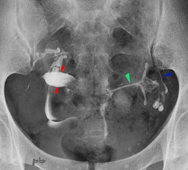
25 – DIU et utérus bicorne
HSG – DIU oublié.
Visualisation d’un DIU pelvien gauche (►). Il est situé dans l’hémicorps utérin gauche. La trompe gauche (►) est bien visible.
Hémicorps droit (►).
25 – IUD and bicornuate uterus
HSG – Forgotten IUD.
Visualization of a left pelvic IUD (►). It is located in the left uterine hemi-cavity. The left fallopian tube (►) is clearly visible.
Right hemi-cavity (►).
© Dr Philippe BASSNAGEL – 2022