
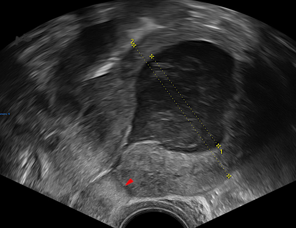
1 – Hématométrie
Rétention hématique après césarienne.
La cavité utérine (croix 1) est élargie et remplie de sang hypoéchogène.
Cicatrice de césarienne (►)
1 – Hematometra
Hematic retention after cesarean section.
The uterine cavity (cross 1) is enlarged and filled with hypoechoic blood.
Cesarean section scar (►).
2 – Hématométrie – Ecoulement
Vidéo-Coupe sagittale échographique sur l’utérus.
Patiente en début de règles.
Ecoulement sanguin bien visible. Il s’effectue du fond utérin (à droite sur la vidéo) vers le col (à gauche).
Les contractions utérines sont également bien visibles au niveau du corps.
Cicatrice de césarienne (►). Remarquez la perte de substance, remplie par le sang.
2 – Hematometra – Discharge
Ultrasound sagittal video section of the uterus.
Patient at the beginning of her period.
Blood flow is clearly visible. It moves from the uterine fundus (right side of the video) toward the cervix (left side).
Uterine contractions are also clearly visible in the uterine body.
Cesarean section scar (►). Note the loss of tissue, filled with blood.
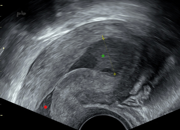
3 – Hématométrie – Sténose de l’orifice externe (OE)
Echographie endovaginale.
Rétention hématique.
La cavité utérine (croix 1) est élargie et remplie de sang hypoéchogène (★).
Il existe aussi une dilation du canal cervical (★) ou hématocolpos.
3 – Hematometra – Stenosis of the External Os
Transvaginal ultrasound.
Hematic retention.
The uterine cavity (cross 1) is enlarged and filled with hypoechoic blood (★).
There is also dilation of the cervical canal (★), or hematocolpos.
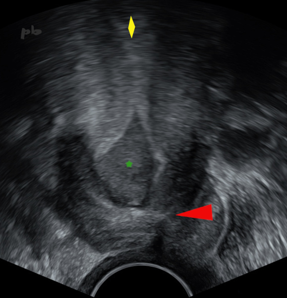
4 – Hématométrie – Sténose de l’orifice externe (OE)
(même patiente que 3)
Echographie endovaginale.
Rétention hématique. Hématocolpos avec un canal cervical très élargi (★) en lien avec une sténose de l’OE du col (►).
Corps utérin (♦)
4 – Hematometra – Stenosis of the external os
(Same patient as case 3)
Transvaginal ultrasound.
Hematic retention. Hematocolpos with a significantly dilated cervical canal (★) due to stenosis of the external os of the cervix (►).
Uterine body (♦).
5 – Hématométrie et hématocolpos
Echographie endovaginale. Vidéo.
Présence de sang au niveau corporéal et cervical sur sténose de l’OE (orifice externe) du col.
5 – Hematometra and hematocolpos
Transvaginal ultrasound. Video.
Presence of blood in the uterine body and cervix due to stenosis of the external os of the cervix.
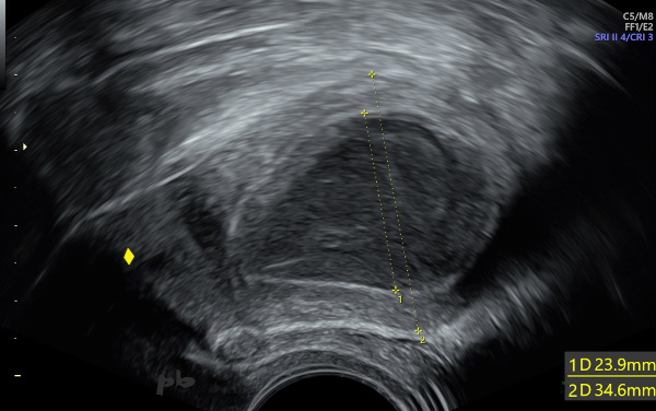
6 – Diagnostic différentiel : pyométrie
Echographie endovaginale.
Métrorragies post-ménopausiques.
Dilatation de la cavité utérine (croix 1) par un liquide hypoéchogène hétérogène.
Le curetage révèlera qu’il s’agissait de pus.
Emdométrite à l’anapath.
Col utérin (♦)
6 – Differential diagnosis: Pyometra
Transvaginal ultrasound.
Postmenopausal metrorrhagia.
Dilation of the uterine cavity (cross 1) with heterogeneous hypoechoic fluid.
Curettage revealed it was pus.
Endometritis confirmed on histopathology.
Cervix (♦).
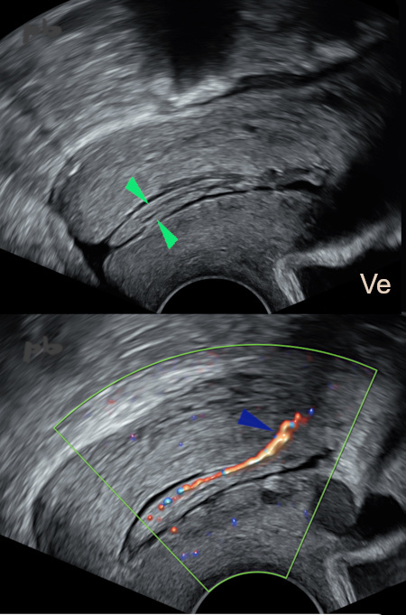
7 – Polype endocervical
Echographie endovaginale – coupes sagittales centrées sur le col.
Visualisation d’une image allongée dans le canal cervical (►).
Le doppler couleur montre le pédicule vasculaire (►), se rattachant à la face postérieure de la région cervico-isthmique.
Ve = vessie
7 – Endocervical polyp
Transvaginal ultrasound – sagittal views centered on the cervix.
Visualization of an elongated lesion in the cervical canal (►).
Color Doppler shows the vascular pedicle (►), attached to the posterior aspect of the cervico-isthmic region.
Ve = bladder
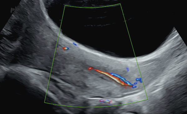
8 – Polype endocervical
Grossesse de 18SA.
Echographie par voie sus-pubienne – coupe sagittale centrée sur le col.
Visualisation d’une image de pédicule vasculaire en doppler couleur dans la lumière cervicale.
8 – Endocervical polyp
18-week pregnancy.
Transabdominal ultrasound – sagittal view centered on the cervix.
Visualization of a vascular pedicle on color Doppler within the cervical lumen.
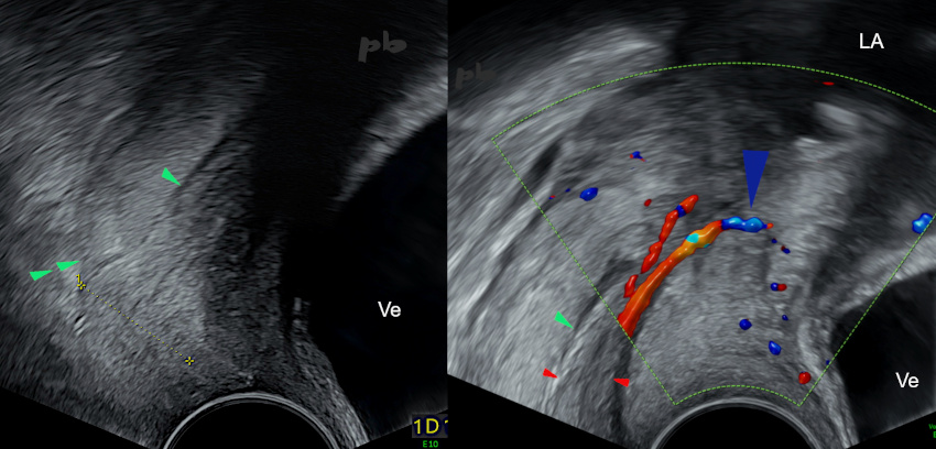
9 – Polype endocervical
(même patiente que 8)
Echographie endovaginale – coupes sagittales centrées sur le col.
- à gauche : polype (entre les croix) accouché par le col. Lèvre postérieure (►►) et face postérieure du canal cervical (►).
- à droite : polype (►) et pédicule (►) s’implantant en avant vers l’orifice interne du col.
Ve = vessie – LA = liquide amniotique
9 – Endocervical polyp (Same patient as case 8)
Transvaginal ultrasound – sagittal views centered on the cervix.
*Left: Polyp (between crosses) protruding through the cervix. Posterior lip (►►) and posterior aspect of the cervical canal (►).
*Right: Polyp (►) and pedicle (►) implanting anteriorly toward the internal os of the cervix.
Ve = bladder – LA = amniotic fluid
© Dr Philippe BASSNAGEL – 2026