
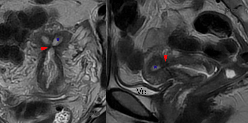
19 – Synéchies
(même patiente que 18)
IRM coupes frontale et sagittale T2 de l’utérus.
Contexte initial d’hémorragie du post-partum traitée par embolisation utérine.
Synéchie (►) en hyposignal T2, fermant la cavité.
Endomètre (★)
Ve = vessie
19 – Synechiae
(Same patient as case 18)
MRI – T2-weighted frontal and sagittal views of the uterus.
Initial context: Postpartum hemorrhage treated with uterine artery embolization.
Synechia (►) appears as a T2 hypointense signal, obliterating the uterine cavity.
Endometrium (★) Ve = Bladder
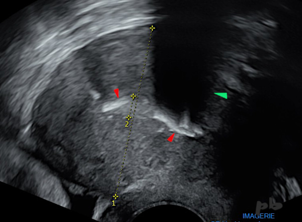
20 – Calcification et synéchie
(même patiente que 16)
Echographie réalisée 9 mois après l’HSG. Antécédent de curetage.
Apparition d’une volumineuse calcification (►) avec cône d’ombre (►).
20 – Calcification and Synechia
(Same patient as case 16)
Ultrasound performed 9 months after the HSG. History of curettage.
Appearance of a large calcification (►) with posterior acoustic shadowing (►).
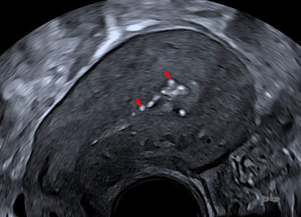
21 – Calcifications
Révision utérine il y a 2 mois.
Echographie endovaginale.
Présence de calcifications (►) postérieures (le placenta était postérieur pendant la grossesse) : simples phénomènes cicatriciels en rapport avec le curetage, ou persistance de quelques résidus placentaires.
21 – Calcifications
Uterine revision performed 2 months ago.
Transvaginal ultrasound.
Presence of posterior calcifications (►) (the placenta was posterior during pregnancy): either simple scar phenomena related to the curettage, or persistence of some placental residues.
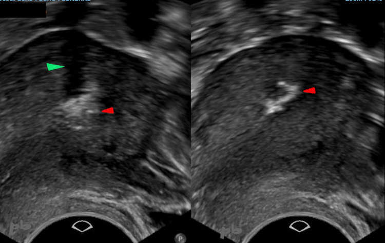
22 – Calcifications
(même patiente que 21)
Révision utérine il y a 4 mois.
Echographie endovaginale.
Progression des calcifications (►) par rapport à l’échographie précédente 2 mois plus tôt.
Cône d’ombre (►)
22 – Calcifications
(Same patient as case 21)
Uterine revision performed 4 months ago.
Transvaginal ultrasound.
Progression of calcifications (►) compared to the previous ultrasound 2 months earlier.
Posterior acoustic shadowing (►).
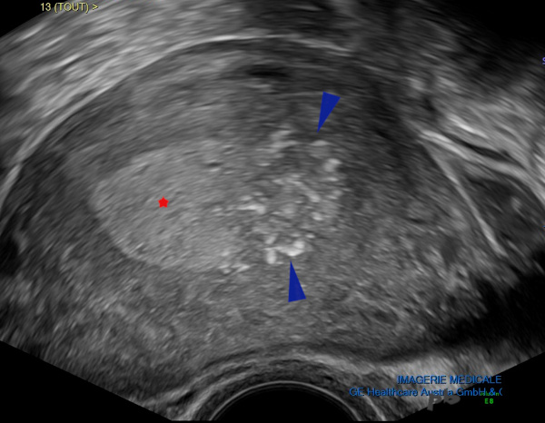
23 – Calcifications placentaires
Accouchement par voie basse il y a 2 mois et demi. Métrorragies.
Persistance d’un résidu placentaire (►) ponctué de petites calcifications.
Endomètre (★).
23 – Placental Calcifications
Vaginal delivery 2.5 months ago. Metrorrhagia.
Persistence of a placental residue (►) dotted with small calcifications.
Endometrium (★).
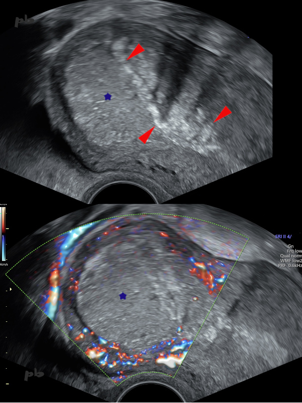
24 – Calcifications sur placenta acreta
Placenta acreta. Résidus placentaires laissés en place lors de l’accouchement (★).
Evolution attendue vers la régression, la dévascularisation (absence de flux en doppler couleur), et la calcification (►).
24 – Calcifications in Placenta Accreta
Placenta accreta. Placental residues left in place during delivery (★).
Expected progression toward regression, devascularization (no flow on color Doppler), and calcification (►).
© Dr Philippe BASSNAGEL – 2026