

13 – Endométriose postérieure – Echographie
Coupe sagittale.
Nodule endométriosique spiculé hypoéchogène (entre les croix) localisé sur le torus et fixé sur un segment recto-sigmoïdien. La musculeuse (►), hypoéchogène, est effacée à ce niveau.
Utérus : col/isthme (★) et corps (★).
13 – Posterior Endometriosis – Ultrasound
Sagittal view.
Hypoechoic spiculated endometriotic nodule (between the crosses) located on the torus and attached to a rectosigmoid segment. The muscularis layer (►), which is hypoechoic, is effaced at this level.
Uterus: cervix/isthmus (★) and body (★).

14 – Endométriose postérieure – IRM
(même patiente que 13)
Coupe sagittale T2.
Nodule endométriosique du torus en continuité avec la face antérieure épaissie du rectosigmoïde (★).
En T1 (non montré), il existe des spots en hypersignal.
Cicatrice d’hystérotomie (►).
14 – Posterior Endometriosis – MRI (Same patient as in 13)
Sagittal T2-weighted image.
Endometriotic nodule of the torus in continuity with the thickened anterior wall of the rectosigmoid (★).
On T1-weighted imaging (not shown), there are hyperintense spots.
Hysterotomy scar (►).

15 – Endométriose postérieure – Echodoppler
Coupe sagittale montrant un nodule endométriosique hypoéchogène (►) fixé sur le torus (★) en avant et un segment recto-sigmoïdien en arrière. Infiltration de la paroi digestive.
La lumière digestive est bien visible (►), soulevée par un petit kyste (►) présentant une image de niveau liquidien, endométriosique.
Musculeuse indemne (★).
15 – Posterior Endometriosis – Echodoppler
Sagittal view showing a hypoechoic endometriotic nodule (►) attached to the torus (★) anteriorly and a rectosigmoid segment posteriorly. Infiltration of the digestive wall.
The digestive lumen is clearly visible (►), elevated by a small cyst (►) presenting a fluid-level image, characteristic of endometriosis.
Muscular layer is intact (★).

16 – Endométriose postérieure – Echographie
Nodule endométriosique hypoéchogène (croix).
Une portion de ce nodule (★) infiltre la paroi sigmoïdienne. La musculeuse est totalement effacée à ce niveau. Elle reste visible ailleurs (►).
16 – Posterior Endometriosis – Ultrasound
Hypoechoic endometriotic nodule (cross).
A portion of this nodule (★) infiltrates the sigmoid wall. The muscular layer is completely effaced at this level. It remains visible elsewhere (►).

17 – Endométriose postérieure – IRM
(même patiente que 16)
Coupe axiale T2.
Nodule endométriosique spiculé (►) venant du torus et du ligament utérosacré droit, infiltrant le rectosigmoïde épaissi (★).
L’importance de la pénétration digestive est plus difficile à évaluer par rapport à l’échographie vaginale.
Ovaires (►).
17 – Posterior Endometriosis – MRI (Same patient as 16)
Axial T2-weighted image.
Spiculated endometriotic nodule (►) originating from the torus and the right uterosacral ligament, infiltrating the thickened rectosigmoid (★).
The extent of digestive penetration is more difficult to assess compared to vaginal ultrasound.
Ovaries (►).
18 – Endométriose postérieure – Echographie
Séquence vidéo dans le plan sagittal.
Endométriose sévère.
Nodule endométriosique sur le torus, hypoéchogène (►), s’étendant en arrière vers le sigmoïde. La musculeuse (★) est infiltrée mais non dépassée. La lumière sigmoïdienne (►) est bien visible par moment, sous la forme d’une ligne hyperéchogène, intacte.
Le tissu endométriosique (►) rejoint l’ovaire droit qui est porteur d’un kyste hémorragique fonctionnel (★), ayant disparu sur une IRM réalisée 2 mois plus tard.
18 – Posterior Endometriosis – Ultrasound
Video sequence in the sagittal plane.
Severe endometriosis.
Endometriotic nodule on the torus, hypoechoic (►), extending posteriorly toward the sigmoid. The muscularis (★) is infiltrated but not breached. The sigmoid lumen (►) is occasionally well visible as a hyperechoic line, intact.
The endometriotic tissue (►) connects to the right ovary, which contains a functional hemorrhagic cyst (★), which disappeared on an MRI performed 2 months later.

19 – Endométriose postérieure – Echographie
Patiente ne se plaignant pas de rectorragies, mais présentant des épisodes sub-occlusifs.
Nodule endométriosique hypoéchogène irrégulier marqué par les croix.
Il envahit la paroi sigmoïdienne. La musculeuse a complètement disparu (►) et le nodule proche, voire dans la lumière digestive.
Musculeuse normale (►).
19 – Posterior Endometriosis – Ultrasound
Patient does not report rectorrhagia but experiences sub-occlusive episodes.
Irregular hypoechoic endometriotic nodule marked by crosses.
It invades the sigmoid wall. The muscularis has completely disappeared (►), and the nodule is near or even within the digestive lumen.
Normal muscularis (►).
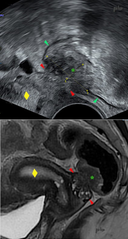
19bis – Endométriose digestive
Echographie endovaginale coupe sagittale (en haut) – IRM coupe sagittale T2 (en bas).
Volumineux nodule endométriosique situé sur le torus (►) et infiltrant en arrière la paroi antérieure du rectosigmoïde.
Cette envahissement (entre les croix – ★) apparait plutôt hypoéchogène et hyposignal. La lumière digestive est respectée en IRM. Interruption de la musculeuse (►) en regard du nodule en échographie.
Utérus (♦)
Cette endométriose profonde n’a été découverte qu’en cours de stimulation ovarienne, alors que les douleurs remontaient à plusieurs années en arrière : ce qui montre la difficulté du diagnostic, et probablement le développement des lésions sous stimulation.
19b – Digestive Endometriosis
Endovaginal ultrasound, sagittal view (top) – MRI, sagittal T2-weighted image (bottom).
Large endometriotic nodule located on the torus (►), infiltrating the anterior wall of the rectosigmoid posteriorly.
This infiltration (between the crosses – ★) appears mostly hypoechoic and hypointense. The digestive lumen is preserved on MRI. Interruption of the muscularis (►) opposite the nodule on ultrasound.
Uterus (♦)
This deep endometriosis was only discovered during ovarian stimulation, even though the pain dated back several years: this highlights the difficulty of diagnosis and likely the progression of lesions under stimulation.
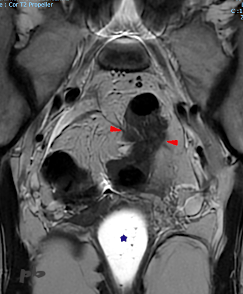
19ter – Endométriose digestive
IRM coupe coronale T2.
Volumineux nodule endométriosique (►) sur le recto-sigmoïde.
Gel intra-vaginal (★)
19c – Digestive Endometriosis
MRI, coronal T2-weighted image.
Large endometriotic nodule (►) on the rectosigmoid.
Intravaginal gel (★)

20 – Endométriose postérieure – Echographie
Coupe sagittale.
Petites lésions endométriosiques vaginales à l’examen clinique.
La sonde endovaginale est positionnée dans le CDS postérieur.
On constate un net épaississement de la paroi vaginale, hypoéchogène (►).
Reproductibilité de la douleur.
Col utérin (★).
20 – Posterior Endometriosis – Ultrasound
Sagittal view.
Small endometriotic lesions in the vagina detected during clinical examination.
The endovaginal probe is positioned in the posterior cul-de-sac.
There is a clear thickening of the vaginal wall, which appears hypoechoic (►).
Pain is reproducible.
Cervix (★).

21 – Endométriose postérieure – IRM
(même patiente que 20)
Coupe axiale T1.
Même aspect d’épaississement de la paroi vaginale, parsemée de multiples images punctiformes en hyper T1 (►) en coupe transversale : atteinte vaginale endométriosique.
Col utérin (★).
21 – Posterior Endometriosis – MRI (Same patient as 20)
Axial T1-weighted image.
Same appearance of vaginal wall thickening, dotted with multiple punctiform hyperintense T1 signals (►) in cross-section: endometriotic vaginal involvement.
Cervix (★).
© Dr Philippe BASSNAGEL – 2022