
22 – Endométriose postérieure – Echographie
Séquence vidéo dans le plan sagittal.
Utérus en minime rétroflexion.
Petite bande hypoéchogène tendue entre le fond vaginal et le ligament utérosacré droit (►)
Douleur élective à la palpation, ce qui a aidé à la repérer.
Probable localisation endométriosique.
Canal cervical (►).
Endomètre (★).
22 – Posterior Endometriosis – Ultrasound
Video sequence in the sagittal plane.
Uterus in slight retroflexion.
Small hypoechoic band stretched between the vaginal fornix and the right uterosacral ligament (►).
Selective pain on palpation, which helped identify it.
Likely endometriotic location.
Cervical canal (►).
Endometrium (★).
23 – Endométriose postérieure – Echo voie sus-pubienne
Coupe sagittale médiane vessie pleine.
Utérus antéversé (entre les croix).
Canal cervical (►).
Image « en plus » en arrière du col, hypoéchogène (►).
23 – Posterior Endometriosis – Suprapubic Ultrasound
Median sagittal view with a full bladder.
Anteverted uterus (between the crosses).
Cervical canal (►).
Additional hypoechoic image behind the cervix (►).

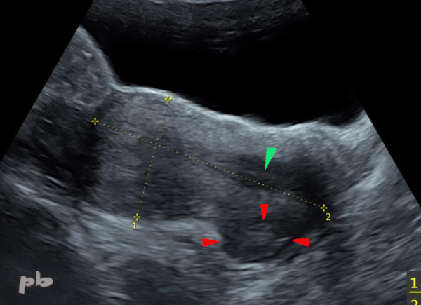
24 – Endométriose postérieure – Echo voie endovaginale
(même patiente que 23)
Coupe sagittale.
Canal cervical (►) en avant. Rectum en arrière (►).
Entre les 2, image hypoéchogène (entre les croix). Elle s’étend sur le ligament utéro-sacré droit et la paroi vaginale du CDS postérieur. La sonde échographique se situe dans le CDS vaginal postérieur, au contact du nodule.
24 – Posterior Endometriosis – Endovaginal Ultrasound (Same patient as 23)
Sagittal view.
Cervical canal (►) in the front. Rectum (►) in the back.
Between the two, a hypoechoic image (between the crosses). It extends to the right uterosacral ligament and the vaginal wall of the posterior cul-de-sac. The ultrasound probe is located in the posterior vaginal cul-de-sac, in contact with the nodule.

25 – Endométriose postérieure – IRM
(même patiente que 23)
Coupe axiale T2.
Canal cervical (►).
On retrouve l’image échographique, située à ce niveau dans la paroi vaginale (►), parsemée de micro kystes en hypersignal.
25 – Posterior Endometriosis – MRI (Same patient as 23)
Axial T2-weighted slice.
Cervical canal (►).
The ultrasound image is identified at this level in the vaginal wall (►), interspersed with microcysts appearing as hyperintense signals.
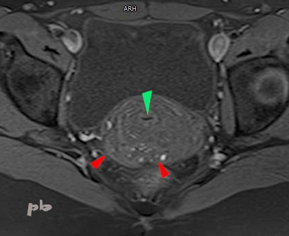
26 – Endométriose postérieure – IRM
(même patiente que 23)
Coupe axiale T1.
Canal cervical (►).
Image hyposignal T1, au même niveau, dans la paroi vaginale (►), parsemée de micro kystes en hypersignal T1 (contenu hématique) : endométriose vaginale.
26 – Posterior Endometriosis – MRI (Same patient as 23)
Axial T1-weighted slice.
Cervical canal (►).
T1-hypointense image, at the same level, in the vaginal wall (►), interspersed with T1-hyperintense microcysts (hemorrhagic content): vaginal endometriosis.
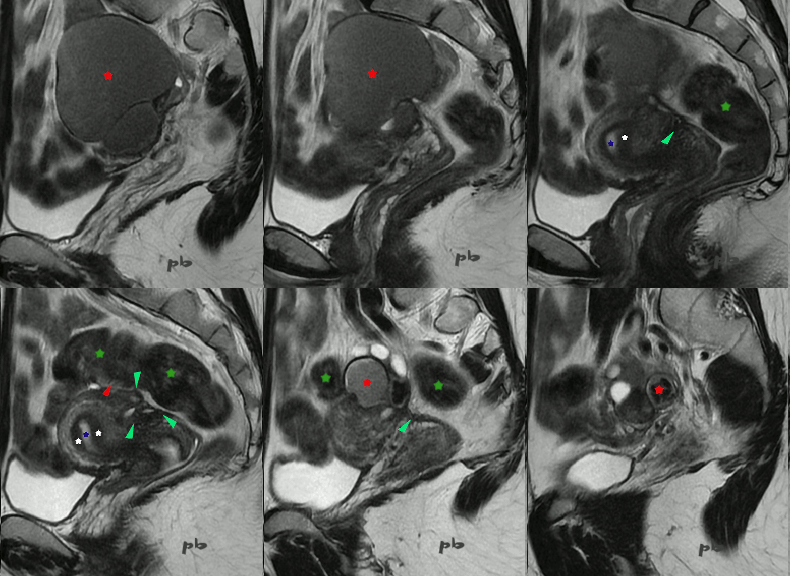
27 – Endométriose postérieure – IRM
Séquence sagittale T2.
Nodule spiculé (►) en hyposignal. Microkystes internes en hypersignal.
Il est situé sur le torus, fixé aux 2 ovaires et au recto sigmoïde (★).
Plusieurs kystes endométriosiques (★) en signal intermédiaire, le plus gros à gauche (sur les 1ères coupes). Sur l’ovaire droit (dernières coupes), un kyste endométriosique avec franc hyposignal (« shading » ★).
La paroi digestive semble épaissie avec une lumière au contraire rétrécie (►) au niveau de la zone d’adhérence.
Net épaississement de la paroi myométriale postérieure, avec plage hyposignal parsemée de kystes : adénomyose externe (►) en continuité avec le nodule endométriosique lui-même.
Endomètre (★).
Zone jonctionnelle (étoile blanche) non épaissie.
27 – Posterior Endometriosis – MRI
Sagittal T2-weighted sequence.
Spiculated nodule (►) with hypointense signal. Internal microcysts with hyperintense signal.
It is located on the torus, attached to both ovaries and the rectosigmoid (★).
Multiple endometriotic cysts (★) with intermediate signal, the largest on the left (on the first slices). On the right ovary (last slices), an endometriotic cyst with marked hypointense signal (« shading » ★).
The digestive wall appears thickened with a conversely narrowed lumen (►) at the adhesion site.
Clear thickening of the posterior myometrial wall, with a hypointense area interspersed with cysts: external adenomyosis (►) in continuity with the endometriotic nodule itself.
Endometrium (★).
Junctional zone (white star) not thickened.

28 – Ligament utéro-sacré – Echographie
Patiente aux antécédents d’endométriome ovarien droit opéré 3 ans auparavant.
Pas de douleur. Sous pilule.
Coupe sagittale par voie endovaginale.
Utérus rétrofléchi. Endomètre (★).
Mise en évidence du ligament utéro-sacré gauche en coupe transversale (entre les croix), modérément épaissi, indolore à la palpation : aspect probablement séquellaire.
Les ligament utéro-sacrés ne sont jamais visualisés à l’état normal en échographie (au contraire de l’IRM).
Ve = vessie
28 – Uterosacral Ligament – Ultrasound
Patient with a history of right ovarian endometrioma surgery 3 years prior.
No pain. On birth control pills.
Sagittal view via endovaginal ultrasound.
Retroflexed uterus. Endometrium (★).
Visualization of the left uterosacral ligament in transverse section (between the crosses), moderately thickened, painless on palpation: likely a sequelae appearance.
Uterosacral ligaments are never visualized in a normal state on ultrasound (unlike MRI).
Ve = Bladder
© Dr Philippe BASSNAGEL – 2022