

13 – Utérus bicorne bicervical (didelphe)
(U3b C2 selon la classification de l’ESHRE)
Echographie par voie sus-pubienne.
Présence de 2 hémicorps utérins droit (★) et gauche (étoile blanche).
La vessie pleine (★) vient s’intercaler entre eux 2, rendant l’incisure fundique utérine (►) évidente, et permettant de différencier utérus bicorne et cloisonné.
13 – Bicornuate bicervical uterus (didelphys) (U3b C2 according to the ESHRE classification)
Suprapubic ultrasound.
Presence of two uterine hemi-bodies right (★) and left (white star). The full bladder (★) is positioned between them, making the uterine fundal notch (►) evident and allowing differentiation between a bicornuate and septate uterus.
14 – Utérus bicorne bicervical (didelphe)
(même patiente que 13)
Echographie endo-vaginale – Séquence vidéo.
Balayage dans le plan frontal du pelvis de l’ensemble de l’utérus en commençant par les 2 cols.
On suit la lumière à droite (►) et à gauche (►) depuis les 2 cols jusqu’aux 2 hémicorps utérins.
Importante divergence des 2 hémi-utérus, avec nette incisure fundique.
14 – Bicornuate bicervical uterus (didelphys)
(same patient as 13)
Transvaginal ultrasound – Video sequence.
Frontal plane scanning of the entire uterus, starting from the two cervices. The lumen is followed on the right (►) and left (►) from the two cervices up to the two uterine hemi-bodies. Significant divergence of the two hemi-uteri, with a clear fundal notch.
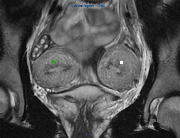
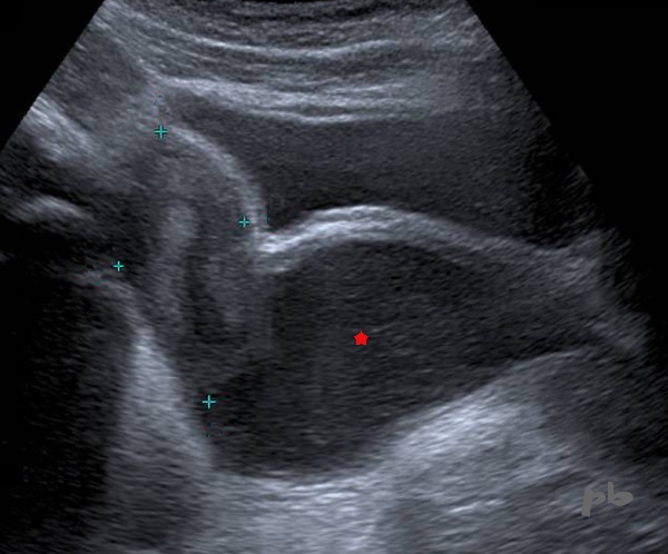
15 – Utérus bicorne bicervical (didelphe)
(même patiente que 13)
IRM coupe frontale T2.
Les 2 hémicorps utérins sont évidents (étoiles), contenant de l’endomètre.
Les ovaires sont aussi visualisés juste au-dessus de leur hémicorps respectif (ils n’appartiennent pas à la malformation au contraire des reins).
15 – Bicornuate bicervical uterus (didelphys)
(same patient as 13)
MRI, frontal T2-weighted section. The two uterine hemi-bodies are evident (stars), containing endometrium. The ovaries are also visualized just above their respective hemi-bodies (they are not part of the malformation, unlike the kidneys).

16 – Utérus bicorne bicervical (didelphe)
(même patiente que 13)
IRM coupe frontale T2.
Les 2 canaux cervicaux sont marqués (étoiles).
En inferieur, les orifices externes sont nettement distants. Mais la visualisation (difficile) des 2 cols est d’abord clinique.
16 – Bicornuate bicervical uterus (didelphys)
(same patient as 13)
MRI, frontal T2-weighted section. The two cervical canals are marked (stars). Inferiorly, the external orifices are clearly distant. However, visualization (which is difficult) of the two cervices is primarily clinical.
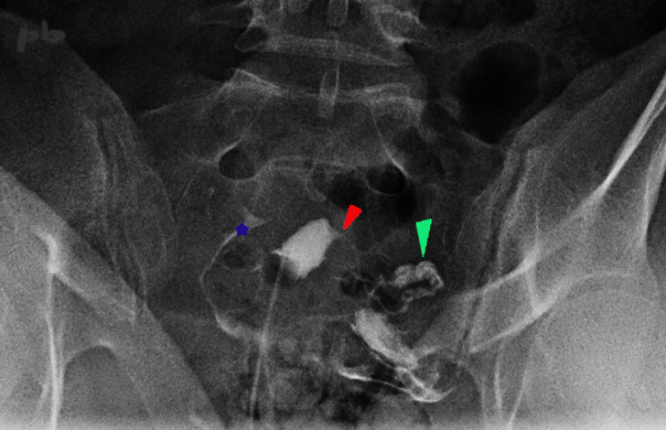
17 – Utérus bicorne bicervical (= utérus didelphe
– U3b C2)
Hystérographie réalisée dans le cadre d’un bilan d’infertilité.
Opacification d’une cavité utérine atypique, et de la trompe gauche (segment interstitiel ► et ampullo-pavillonnaire ►).
Produit de contraste dans la cavité péritonéale du côté droit (★).
Une échographie est donc pratiquée dans les suites, révélant 2 hémicorps utérins ainsi que 2 cols.
L’examen clinique retrouvera alors une cloison vaginale et un 2ème col, à droite, non connu.
Le bilan rénal montre une atrophie à droite, en fait connue depuis plusieurs années.
Le réflexe « malformation utérine -> regarder les reins » doit être complété par le réflexe inverse « malformation rénale -> regarder l’appareil génital ».
17 – Bicornuate bicervical uterus (= didelphic uterus – U3b C2)
Hysterosalpingography performed as part of an infertility assessment.
Opacification of an atypical uterine cavity and the left fallopian tube (interstitial segment ► and ampullary-infundibular segment ►). Contrast medium into the peritoneal cavity on the right side (★). An ultrasound is therefore performed, revealing two uterine hemi-bodies and two cervices. The clinical examination then identifies a vaginal septum and a second, previously unknown cervix on the right. The renal assessment shows atrophy on the right side, which has actually been known for several years.
The reflex “uterine malformation → check the kidneys” should be complemented by the reverse reflex: “renal malformation → check the genital tract.
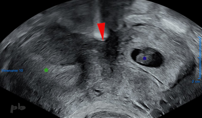
18 – Utérus bicorne bicervical (= didelphe)
(même patiente que 17)
Echographie réalisée 4 ans plus tard, montrant les 2 hémicorps utérins.
Sac gestationnel et embryon de 8SA à gauche (★).
Endomètre (★).
Incisure fundique (►)
Cette patiente avait déjà eu un enfant à 36SA, développé dans l’hémicorps droit.
18 – Bicornuate bicervical uterus (= didelphic uterus)
(same patient as 17)
Ultrasound performed 4 years later, showing the two uterine hemi-bodies. Gestational sac and 8-week embryo on the left (★).
Endometrium (★). Fundal notch (►).
This patient had already given birth to a child at 36 weeks, who developed in the right hemi-body.

19 – Utérus bicorne unicervical en cours de grossesse
(U3b C0 selon la classification de l’ESHRE)
Echographie à 22SA.
Visualisation d’un hémicorps utérin droit (►).
A ne pas confondre avec une masse utérine ou ovarienne si la malformation n’est pas connue.
Endomètre entre les croix.
Membres fœtaux dans l’hémicorps utérin gauche (►).
Liquide amniotique (★).
On peut comparer avec l’image 11 montrant une grossesse sur un utérus cloisonné.
19 – Bicornuate unicervical uterus during pregnancy (U3b C0 according to the ESHRE classification)
Ultrasound at 22 weeks of gestation. Visualization of the right uterine hemi-body (►). Not to be confused with a uterine or ovarian mass if the malformation is unknown. Endometrium between the crosses. Fetal limbs in the left uterine hemi-body (►). Amniotic fluid (★). This can be compared with image 11, showing a pregnancy in a septate uterus.
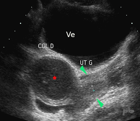
20 – Utérus bicorne avec hématométrie droite
(U3b C2 selon la classification de l’ESHRE)
Voie abdominale chez une jeune fille de 13 ans pour des douleurs cycliques.
Coupe transversale du pelvis.
Distension du col droit (★) par du sang. La paroi est épaisse à ce niveau de coupe, excluant a priori l’origine vaginale.
Aspect normal de l’hémicavité gauche (►).
Le vagin n’était pas dilaté.
Ve = vessie.
20 – Bicornuate uterus with right-sided hematometra
(U3b C2 according to the ESHRE classification)
Abdominal approach in a 13-year-old girl for cyclic pain.
Transverse section of the pelvis.
Distension of the right cervix (★) with blood. The wall is thick at this section level, initially ruling out a vaginal origin.
Normal appearance of the left hemicavity (►).
The vagina was not dilated.
Ve = bladder.
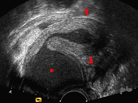
21 – Utérus bicorne avec hématométrie droite
(U3b C2 selon la classification de l’ESHRE)
(même patiente que 20)
Voie endorectale (avec l’accord de la mère et de la jeune fille et préparation psychologique – dossier ancien).
Coupe sagittale sur la cavité droite.
Distension hématique du col droit (★), et de l’hémicavité droite (►).
Paroi myométriale bien visible.
21 – Bicornuate uterus with right-sided hematometra
(U3b C2 according to the ESHRE classification)
(Same patient as image 20)
Endorectal approach (with the consent of the mother and the young girl, and psychological preparation – old medical record).
Sagittal section of the right cavity.
Hematic distension of the right cervix (★) and the right hemicavity (►).
Myometrial wall clearly visible.
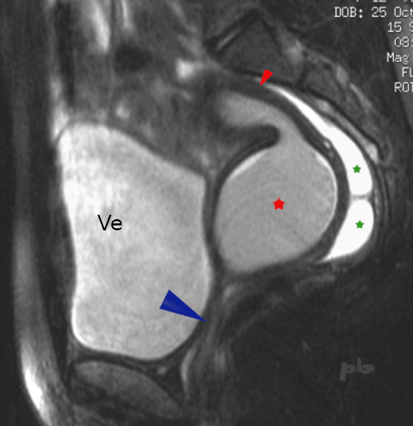
22 – Utérus bicorne avec hématométrie droite
(même patiente que 20)
IRM T2 fat sat réalisée à la même époque. Coupe sagittale sur la cavité droite.
Distension du col droit (★), et de l’hémicavité droite (►).
Le vagin n’est pas distendu (►).
Il existe un épanchement en rapport avec le reflux menstruel tubaire (★) du côté droit obstrué.
Ve = vessie.
22 – Bicornuate uterus with right hematometra
(Same patient as 20)
T2-weighted fat-saturated MRI performed at the same time. Sagittal view of the right cavity.
Distension of the right cervix (★) and the right hemicavity (►). The vagina is not distended (►). There is fluid accumulation due to tubal menstrual reflux (★) on the obstructed right side.
Ve = bladder.
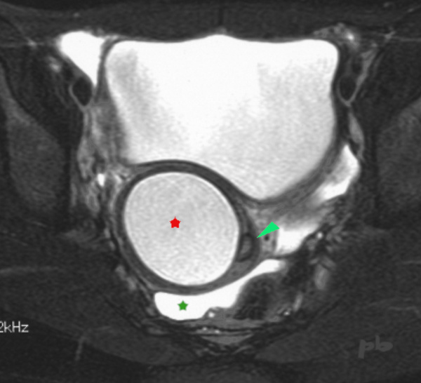
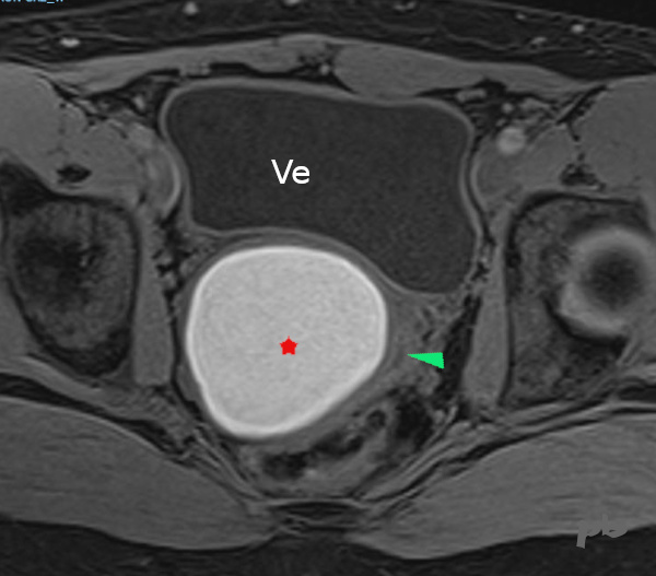
23 – Utérus bicorne avec hématométrie droite
(même patiente que 20)
IRM T2 fat sat réalisée à la même époque. Coupe transversale sur les 2 cols.
Distension du col droit (★).
Col gauche refoulé et comprimé, non dilaté (►).
Epanchement lié au reflux menstruel (★). Il existe par ailleurs une agénésie rénale droite, du côté de la dilatation comme habituellement rencontré.
23 – Bicornuate uterus with right hematometra
(Same patient as 20)
T2-weighted fat-saturated MRI performed at the same time.
Transverse view of both cervices. Distension of the right cervix (★). Left cervix displaced and compressed, not dilated (►). Fluid accumulation due to menstrual reflux (★). Additionally, there is right renal agenesis on the same side as the dilation, as commonly observed.

24 – Utérus didelphe
(U3b C2 selon la classification de l’ESHRE)
(même patiente que 20)
Voie endovaginale. Coupe frontale du pelvis.
12 années plus tard, et après chirurgie, utérus bicorne bicervical classique.
Endomètre de l’hémicavité droite (★).
Endomètre de l’hémicavité gauche (★).
Cette patiente est revue 20 ans après le bilan initial pour une prise charge en AMP.
24 – Didelphic uterus (ESHRE classification U3b C2)
(Same patient as 20)
Endovaginal approach. Frontal view of the pelvis.
Twelve years later, and after surgery, a classic bicornuate bicervical uterus is observed. Endometrium of the right hemicavity (★). Endometrium of the left hemicavity (★). This patient is reviewed 20 years after the initial assessment for assisted reproductive technology (ART) management.
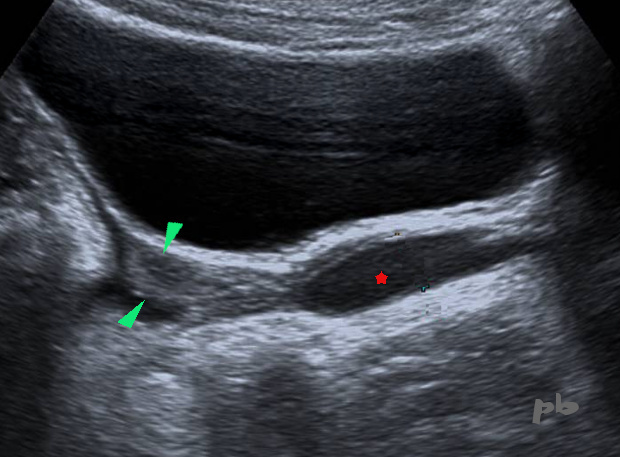
25 – Utérus didelphe avec hématocolpos à droite
(U3b C2 V2 selon la classification de l’ESHRE)
Voie abdominale chez une jeune fille de 11 ans. Bilan dans le cadre d’un suivi pour dysplasie multikystique à droite.
Coupe sagittale.
Distension de l’hémivagin droit (★). Il est surmonté par l’hémi-utérus droit encore de type impubère (►).
25 – Didelphic uterus with right hematocolpos (ESHRE classification U3b C2 V2)
Abdominal approach in an 11-year-old girl. Assessment as part of follow-up for right multicystic dysplasia. Sagittal view.
Distension of the right hemivagina (★). It is topped by the right hemiuterus, still of prepubertal type (►).
26 – Utérus didelphe avec hématocolpos à droite
(U3b C2 V2 selon la classification de l’ESHRE)
(même patiente que 25)
Voie abdominale 3 ans plus tard.
Coupe sagittale.
Distension échogène, d’allure hématique, majorée de l’hémivagin droit (★). Il est surmonté par l’hémi-utérus droit (entre les croix) qui s’est nettement développé.
26 – Didelphic uterus with right hematocolpos (ESHRE classification U3b C2 V2)
(Same patient as 25)
Abdominal approach 3 years later. Sagittal view.
Echoic distension, likely hematometric, increased in the right hemivagina (★). It is topped by the right hemiuterus (between the crosses), which has developed significantly.
27 – Utérus didelphe avec hématocolpos à droite
(même patiente que 25)
IRM 3 ans plus tard.
Coupe axiale T1 fatsat.
Distension de l’hémivagin droit (★) en hypersignal T1.
La lumière de l’hémivagin gauche (►) est visualisée sous la forme d’une image linéaire en hyposignal.
Ve = vessie.
27 – Didelphic uterus with right hematocolpos (Same patient as 25)
MRI 3 years later. Axial T1 fat-saturated view.
Distension of the right hemivagina (★) showing T1 hyperintensity. The lumen of the left hemivagina (►) is visualized as a linear hypointense image.
Ve = bladder.
© Dr Philippe BASSNAGEL – 2022