

1 – Endométriose postérieure – Echographie sus-pubienne
Coupe transversale par voie abdominale vessie pleine.
En arrière du col utérin (★), image hypoéchogène (►) à grand axe antéro-postérieur, de contours anfractueux.
Il est rare de voir aussi bien un nodule du torus par voie abdominale, même vessie pleine.
1 – Posterior endometriosis – Suprapubic ultrasound
Transverse abdominal scan with a full bladder.
Behind the uterine cervix (★), a hypoechoic image (►) with a large anteroposterior axis and irregular contours.
It is rare to visualize a torus nodule so clearly via the abdominal route, even with a full bladder.
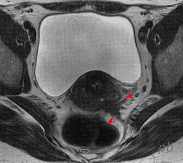
2 -Endométriose postérieure – IRM
Coupe axiale T2.
Aspect typique de nodule spiculé endométriosique en hyposignal (►) situé sur le ligament utérosacré gauche.
Le ligament reprend une morphologie normale plus en arrière. Le droit n’est pas visible sur cette coupe.
2 – Posterior Endometriosis – MRI
Axial T2-weighted image.
Typical appearance of a spiculated endometriotic nodule in hypointense signal (►) located on the left uterosacral ligament.
The ligament regains a normal morphology further posteriorly. The right ligament is not visible on this slice.
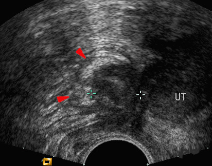
3 – Endométriose postérieure – Echographie endovaginale
Coupe sagittale.
En arrière du col et de l’isthme utérin (UT), image hypoéchogène nodulaire (entre les croix), fixée aux structures digestives en arrière (►). Elle est spiculée, un peu à la manière d’un carcinome mammaire.
Image ancienne mais très démonstrative.
3 – Posterior Endometriosis – Endovaginal Ultrasound
Sagittal view.
Behind the cervix and uterine isthmus (UT), a hypoechoic nodular image (between the crosses), fixed to the digestive structures posteriorly (►). It is spiculated, somewhat resembling a breast carcinoma.
An older image but highly demonstrative.
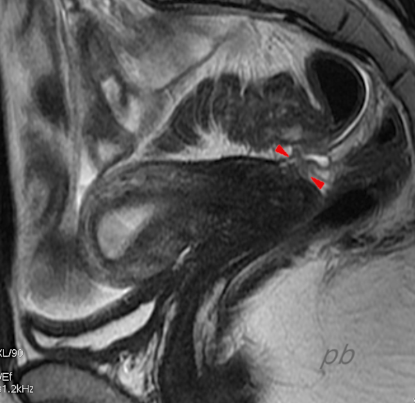
4 – Endométriose postérieure – IRM
Coupe sagittale T2.
Utérus antéversé antéfléchi.
Nodule endométriosique hyposignal (►) tendu entre le torus et le sigmoïde, interrompant le liseré graisseux en hypersignal qui souligne habituellement la face postérieure de l’utérus.
4 – Posterior Endometriosis – MRI
Sagittal T2-weighted image.
Anteverted and anteflexed uterus.
Hypointense endometriotic nodule (►) stretched between the torus and the sigmoid colon, disrupting the hyperintense fatty stripe that typically outlines the posterior surface of the uterus.

5 – Endométriose postérieure – IRM
Coupe axiale T1.
Image hyposignal (►), comprenant plusieurs petits kystes en hypersignal (de nature hématique).
Elle se situe sur le CDS vaginal postérieur et le ligament utérosacré droit.
ligament utérosacré gauche normal (►).
Corps utérin (♦).
5 – Posterior Endometriosis – MRI
Axial T1-weighted image.
Hypointense image (►), containing several small hyperintense cysts (of hemorrhagic nature).
It is located on the posterior vaginal fornix and the right uterosacral ligament.
Left uterosacral ligament appears normal (►).
Uterine body (♦).
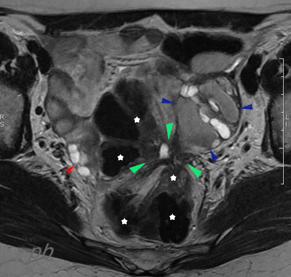
6 – Endométriose postérieure – IRM
Coupe axiale T2.
Image rétractile en hyposignal (►), centrée par une petite zone liquidienne.
On voit le sigmoïde (étoile blanche) et l’ovaire gauche (►) converger sur cette image : aspect stellaire.
L’ovaire gauche est porteur de follicules (en hypersignal) et de kystes endométriosiques (en signal intermédiaire).
Ovaire droit (►).
6 – Posterior Endometriosis – MRI
Axial T2-weighted image.
Retractile hypointense image (►), centered by a small fluid-filled area.
The sigmoid colon (white star) and the left ovary (►) are seen converging on this image: a stellate appearance.
The left ovary contains follicles (hyperintense) and endometriotic cysts (intermediate signal).
Right ovary (►).
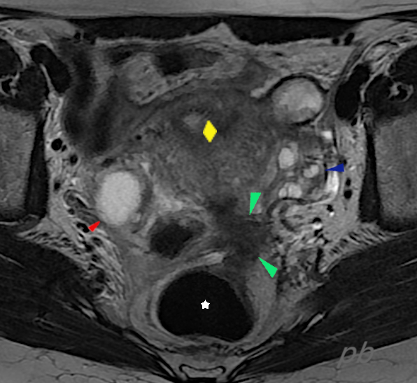
7 – Endométriose postérieure – IRM
(même patiente que 6)
Coupe axiale T2.
Image rétractile en hyposignal (►), mieux individualisée.
On voit toujours le sigmoïde (étoile blanche) et l’ovaire gauche (►) converger sur cette image, qui est d’autre part fixée à l’utérus (♦).
La paroi du sigmoïde apparait épaissie, mais la profondeur de l’infiltration ne peut être évaluée en IRM.
L’ovaire gauche est porteur de follicules (en hypersignal).
Ovaire droit (►).
7 – Posterior Endometriosis – MRI (Same patient as 6)
Axial T2-weighted image.
Retractile hypointense lesion (►), more clearly defined.
The sigmoid colon (white star) and the left ovary (►) are still visible converging on this image, which is also fixed to the uterus (♦).
The sigmoid wall appears thickened, but the depth of infiltration cannot be assessed on MRI.
The left ovary contains follicles (hyperintense).
Right ovary (►).
8 – Endométriose postérieure – Echographie
Séquence vidéo dans le plan frontal du pelvis.
L’examen est demandé en raison de douleurs invalidantes, 5 mois après la réalisation d’une IRM pelvienne considérée initialement comme normale (image 9).
On visualise très bien une petite bande hypoéchogène (►) tendue entre le fond vaginal, le sigmoïde en arrière (★), et probablement située sur le ligament utérosacré gauche.
L’ovaire gauche semble fixé dessus (★).
La douleur est parfaitement reproduite.
Endomètre (grande étoile ★).
8 – Posterior Endometriosis – Ultrasound
Video sequence in the frontal plane of the pelvis.
The examination is requested due to disabling pain, 5 months after a pelvic MRI was performed and initially considered normal (image 9).
A small hypoechoic band (►) is clearly visible, stretched between the vaginal fornix, the sigmoid colon posteriorly (★), and likely located on the left uterosacral ligament.
The left ovary appears fixed to it (★).
The pain is perfectly reproduced.
Endometrium (large star ★).
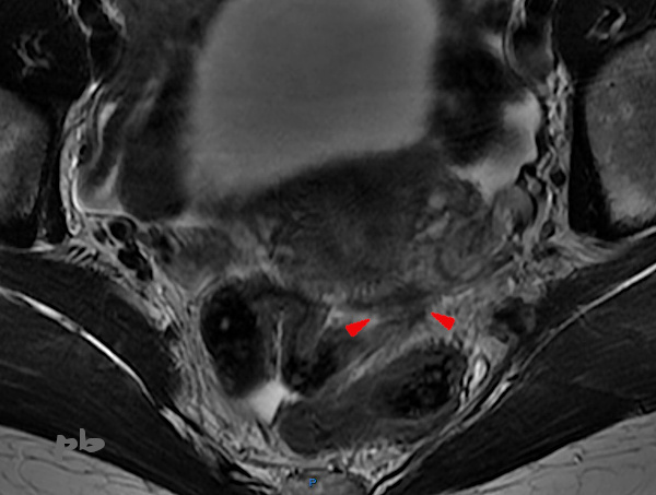
9 – Endométriose postérieure – IRM
(même patiente que 8)
Coupe axiale T2.
En arrière de l’isthme utérin, image hyposignal nodulaire et spiculée (►), sur le torus et le ligament utérosacré gauche.
Cette image n’avait pas été vu lors de la 1ère lecture, et c’est la découverte d’anomalies à l’échographie 5 mois plus tard qui ont amené à redemander une 2ème lecture.
9 – Posterior Endometriosis – MRI (Same patient as 8)
Axial T2-weighted image.
Behind the uterine isthmus, a nodular and spiculated hypointense lesion (►) is visible on the torus and the left uterosacral ligament.
This lesion was not identified during the initial reading, and the discovery of abnormalities on ultrasound 5 months later led to a request for a second review.

10 – Endométriose postérieure – Echographie endovaginale
Coupe sagittale.
En arrière de l’isthme utérin, image hypoéchogène nodulaire (entre les croix).
Cette lésion est plus difficile à repérer en imagerie (écho, IRM) du fait de la rétroflexion utérine.
La reproductibilité de la douleur en échographie est d’une aide précieuse.
Contours utérins (►).
Fond utérin (►).
Endomètre (♦).
10 – Posterior Endometriosis – Endovaginal Ultrasound
Sagittal view.
Behind the uterine isthmus, a nodular hypoechoic lesion (between the crosses).
This lesion is more difficult to detect on imaging (ultrasound, MRI) due to the uterine retroflexion.
The reproducibility of pain during ultrasound is a valuable aid.
Uterine contours (►).
Uterine fundus (►).
Endometrium (♦).
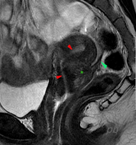
11 – Endométriose postérieure – IRM
Coupe sagittale T2.
En arrière de l’isthme utérin, image hypoéchogène nodulaire (★).
Cette lésion est plus difficile à repérer en imagerie (écho, IRM) du fait de la rétroflexion utérine. Il existe ici un comblement de l’espace graisseux situé normalement dans l’angle col-corps en arrière.
Extension de l’endométriose sur un ligament utérosacré (►).
Canal cervical et endomètre (►).
11 – Posterior Endometriosis – MRI
Sagittal T2-weighted image.
Behind the uterine isthmus, a nodular hypoechoic lesion (★).
This lesion is more difficult to detect on imaging (ultrasound, MRI) due to uterine retroflexion. There is also an obliteration of the fatty space normally located in the cervix-corpus angle posteriorly.
Extension of endometriosis to a uterosacral ligament (►).
Cervical canal and endometrium (►).
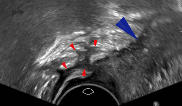
12 – Endométriose postérieure – Echographie endovaginale
Coupe sagittale.
En arrière du col et de l’isthme utérins (►), image hypoéchogène (►) de contours anfractueux, d’allure infiltrante.
Douleur modérée en regard.
Aspect compatible avec une endométriose du torus et des ligaments utérosacrés.
Il s’agit d’une patiente ménopausée sans THS. Pas d’endométriose connue ni d’antécédents chirurgicaux (des adhérences sans tissu endométriosique peuvent aussi être évoquées sur ces images).
12 – Posterior Endometriosis – Endovaginal Ultrasound
Sagittal view.
Behind the cervix and uterine isthmus (►), a hypoechoic image (►) with irregular, infiltrative contours.
Moderate pain at the corresponding site.
Appearance consistent with endometriosis of the torus and uterosacral ligaments.
This is a menopausal patient without hormone replacement therapy. No known endometriosis or surgical history (adhesions without endometriotic tissue may also be considered based on these images).
© Dr Philippe BASSNAGEL – 2022