
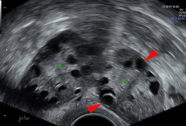
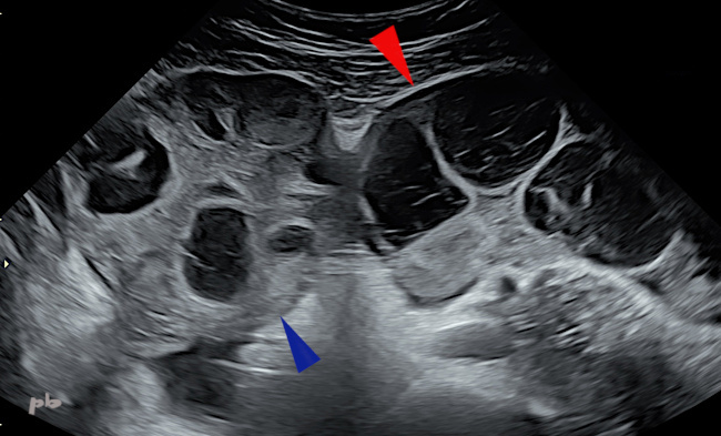
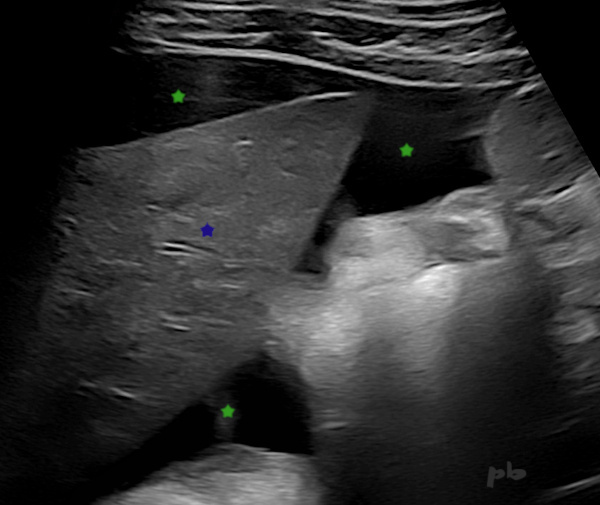
1 – Ovaires micropolykystiques (OPK)
Echographie endovaginale.
2 gros ovaires dans le cul-de-sac de Douglas, en contact l’un avec l’autre (« kissing ovaries »).
Très nombreux petits follicules.
On retrouve 2 aspects morphologiques caractéristiques (mais pas toujours présents) :
-la disposition périphérique des follicules (►)
-l’hypertrophie centrale du stroma (★).
1 – Polycystic Ovary Syndrome (PCOS)
Transvaginal ultrasound.
Two enlarged ovaries located in the pouch of Douglas, in contact with each other (« kissing ovaries »).
Numerous small follicles.
Two characteristic morphological features (though not always present) are observed:
*peripheral arrangement of follicles (►)
*central stromal hypertrophy (★).
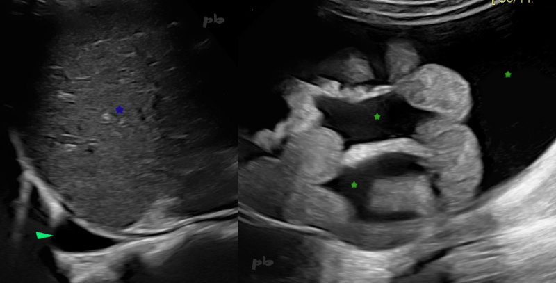
2 – Ovaires micropolykystiques (OPK)
Echographie endovaginale. Séquence vidéo.
Très nombreux follicules, largement supérieurs à 20 (seuil retenu par définition).
2 – Polycystic Ovaries (PCOS)
Transvaginal ultrasound. Video sequence.
Very numerous follicles, well over 20 (the threshold defined for diagnosis).

3 – Ovaire micropolykystique
Echographie endovaginale 3D avec reconnaissance automatique des follicules.
Patiente SOPK (syndrome des ovaires micropolykystiques).
Ovaire porteur d’au moins 52 follicules après stimulation ovarienne (qui a été naturellement rapidement interrompue).
3 – Polycystic Ovary
3D transvaginal ultrasound with automatic follicle recognition.
Patient with PCOS (polycystic ovary syndrome).
Ovary containing at least 52 follicles after ovarian stimulation (which was naturally quickly discontinued).
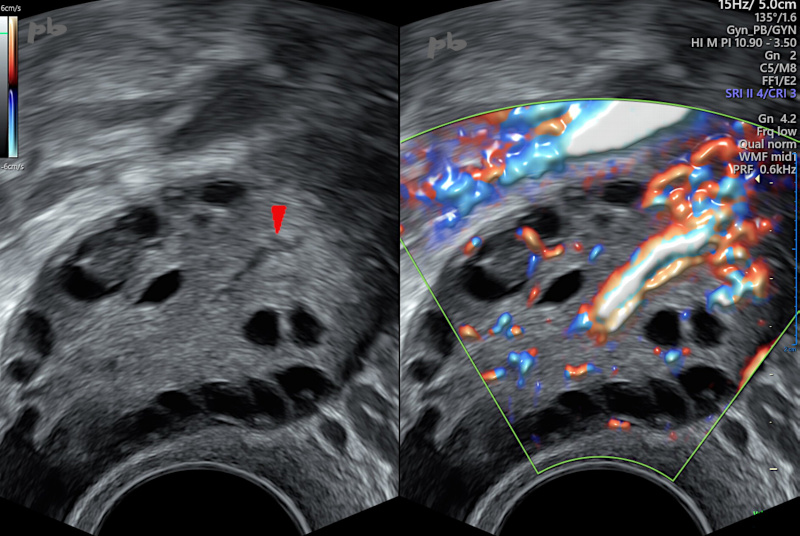
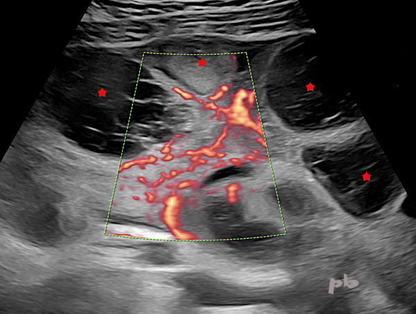
3bis – OPK
Doppler couleur.
Examen réalisé à J3 du cycle.
Il existe une riche vascularisation, habituelle dans ce contexte.
Un vaisseau est même visible en mode B (image gauche ►), ce qui est beaucoup plus rare, mais témoigne de cette hypervascularisation.
3bis – Polycystic ovary
Color Doppler ultrasound.
Exam performed on day 3 of the cycle.
There is rich vascularization, which is usual in this context.
A vessel is even visible in B-mode (left image ►), which is much rarer but indicates this hypervascularization.

4 – Ovaires micropolykystiques (OPK)
IRM coupe axiale T2, montrant la même sémiologie.
La disposition périphérique des follicules (►) et l’hypertrophie centrale du stroma (★) sont évidentes, en plus de leur très grand nombre.
4 – Polycystic Ovaries (PCOS)
Axial T2-weighted MRI scan, showing the same semiology.
The peripheral arrangement of follicles (►) and central stromal hypertrophy (★) are evident, in addition to their very large number.
5 – Hyperstimulation ovarienne
Echographie par voie abdominale.
Présence de 2 volumineux ovaires en fosse iliaque, porteurs de plusieurs images liquidiennes hypoéchogènes hémorragiques.
L’examen est réalisé après la ponction folliculaire (voir aussi chapitre « Endométriome » images 17 et 18 pour les aspects IRM post ponction folliculaire).
Ovaire droit (►)
Ovaire gauche (►)
5 – Ovarian Hyperstimulation
Abdominal ultrasound.
Presence of two significantly enlarged ovaries in the iliac fossa, containing multiple hypoechoic hemorrhagic cystic images.
The examination is performed after follicular puncture (see also the chapter « Endometrioma, » images 17 and 18, for MRI aspects post-follicular puncture).
Right ovary (►)
Left ovary (►)
6 – Hyperstimulation ovarienne
(même patiente que 5)
Echographie par voie abdominale avec doppler.
Volumineux ovaire en fosse iliaque, porteur de plusieurs images liquidiennes hypoéchogènes hémorragiques (★).
Importante vascularisation en doppler couleur, habituelle dans ce contexte.
6 – Ovarian hyperstimulation
(same patient as 5)
Abdominal ultrasound with Doppler.
Large ovary in the iliac fossa, containing multiple hypoechoic hemorrhagic cystic images (★).
Significant vascularization on color Doppler, typical in this context.
7 – Hyperstimulation ovarienne
(même patiente que 5)
Echographie par voie abdominale.
Epanchement péri-hépatique marqué (★).
La présence d’un épanchement péritonéal correspond à une hyperstimulation de grade 3 dans la classification de Nolan modifié (sur 5). Il faut aussi le rechercher en pelvien et dans les gouttières pariéto-coliques. Le rechercher aussi en pleural.
Foie (★)
7 – Ovarian hyperstimulation
(same patient as 5)
Abdominal ultrasound.
Marked perihepatic effusion (★).
The presence of peritoneal effusion corresponds to grade 3 hyperstimulation in the modified Nolan classification (out of 5). It should also be investigated in the pelvic region and in the parietocolic gutters. It should also be checked for pleural effusion.
Liver (★)

8 – Hyperstimulation ovarienne
Acquisition volumique 3D avec reconstruction du volume, et comptage folliculaire.
Au moins 39 follicules sont ainsi dénombrés sur cet ovaire.
Patiente suivie en Assistance Médicale à la Procréation pour des ovaires micropolykystiques, facteur de risque d’hyperstimulation.
8 – Ovarian hyperstimulation
3D volumetric acquisition with volume reconstruction and follicular count.
At least 39 follicles are counted on this ovary.
The patient is being monitored in Assisted Reproductive Technology for micropolycystic ovaries, a risk factor for hyperstimulation.

9 – Hyperstimulation ovarienne
Echographie. Coupe transversale sur le pelvis par voie sus-pubienne.
L’utérus au centre de l’image (♦) semble flotter dans le liquide péritonéal (★), seulement rattaché à la paroi par les ligaments larges (►).
9 – Ovarian hyperstimulation
Ultrasound. Transverse section of the pelvis via the suprapubic route.
The uterus in the center of the image (♦) appears to float in the peritoneal fluid (★), only attached to the wall by the broad ligaments (►).
9bis – Epanchement pleural et péritonéal
Echographie.
Hyperstimulation ovarienne.
Epanchement pleural droit (►) et péritonéal (★) silhouettant les anses digestives et le mésentère.
Foie (★)
9bis – Pleural and peritoneal effusion- Ultrasound
Ovarian hyperstimulation.
Right pleural effusion (►) and peritoneal effusion (★) outlining the bowel loops and mesentery.
Liver (★)
© Dr Philippe BASSNAGEL – 2024