
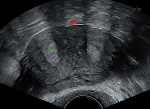
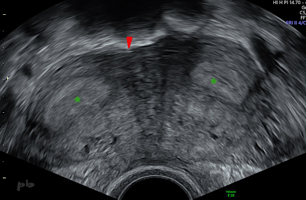
1 – Utérus cloisonné total
(U2b C1 selon la classification de l’ESHRE)
Echographie endo-vaginale. Coupe transversale sur les 2 hémicorps utérins.
Endomètre (★).
Absence d’incisure fundique (►), excluant un utérus bicorne (U3).
1 – Total septate uterus (ESHRE classification : U2b C1)
Transvaginal ultrasound. Transverse section showing both uterine hemi-cavities.
Endometrium (★).
Absence of a fundal notch (►), ruling out a bicornuate uterus (U3).
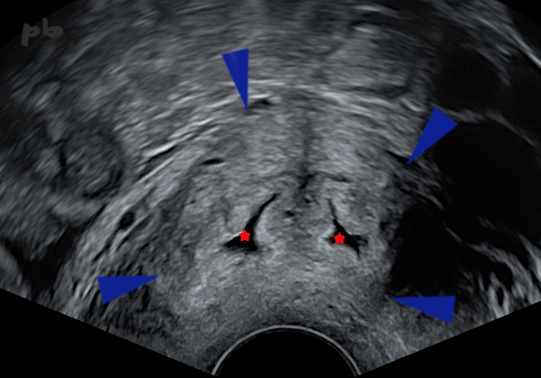
2 – Utérus cloisonné total
(même patiente que 1)
Echographie endo-vaginale. Coupe transversale sur le col.
Un seul massif cervical (►), et 2 canaux cervicaux (★) séparés par une cloison.
2 – Total septate uterus
(same patient as image 1)
Transvaginal ultrasound. Transverse section of the cervix.
Single cervical mass (►), with two separate cervical canals (★) divided by a septum.
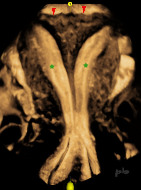
3 – Utérus cloisonné total
(même patiente que 1)
Echographie endo-vaginale et reconstruction frontale après acquisition volumique.
Les 2 hémi-cavités (★) sont suivies du fond utérin jusqu’à l’orifice externe cervical : vue « hystérographique ».
On visualise bien en haut l’aspect rectiligne et aplati du contour externe du fond (►), et l’absence d’incisure fundique.
3 – Complete septate uterus
(same patient as image 1)
Transvaginal ultrasound with frontal reconstruction after volumetric acquisition. Both hemi-cavities (★) are visualized from the uterine fundus to the external cervical os : a « hysterosalpingographic » view. The straight, flattened appearance of the external fundal contour (►) and the absence of a fundal notch are clearly visible at the top.
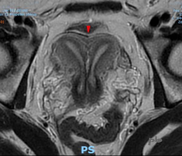
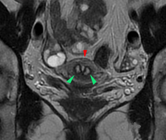
4 – Utérus cloisonné total
(U2b C1 selon la classification de l’ESHRE)
IRM. Coupe transversale du pelvis et coupe frontale de l’utérus.
Visualisation complète de 2 hémicorps utérins.
Discrète incisure fundique (►), inférieure à 50 % de l’épaisseur du myomètre et donc ne permettant pas de classer cet utérus comme bicorne (U3).
4 – Complete septate uterus (ESHRE classification: U2b C1)
MRI. Transverse pelvic section and frontal uterine section. Complete visualization of two uterine hemi-cavities. Slight fundal indentation (►), less than 50% of the myometrial thickness, thus not meeting the criteria for a bicornuate uterus (U3).
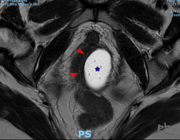
5 – Utérus cloisonné total
(U2b C1 selon la classification de l’ESHRE)
(même patiente que 4)
IRM. Coupe frontale du pelvis et coupe transversale de l’utérus.
Un seul massif cervical (►), et 2 canaux cervicaux (►).
5 – Complete septate uterus
(ESHRE classification: U2b C1)
(same patient as image 4)
MRI. Frontal pelvic section and transverse uterine section.
Single cervical mass (►), with two cervical canals (►).
6 – Utérus cloisonné total
(U2b C1 selon la classification de l’ESHRE)
(même patiente que 4)
IRM coupe axiale au niveau vaginal, après mise en place de gel intra-vaginal par la patiente.
Hémi vagin gauche rempli par le gel échographique (★).
Hémi vagin droit repéré grâce à sa lumière en hypersignal T2 (►).
La cloison vaginale est refoulée vers la droite par le gel.
6 – Complete septate uterus (U2b C1 according to ESHRE classification)
(same patient as 4)
Axial MRI at the vaginal level after the patient inserted intravaginal gel. The left hemi-vagina is filled with ultrasound gel (★). The right hemi-vagina is identified by its T2 hyperintense lumen (►). The vaginal septum is displaced to the right by the gel.
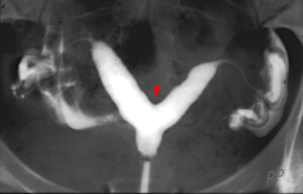
7 – Utérus cloisonné partiel
(U2a C0 selon la classification de l’ESHRE)
Hystérographie (HSG).
2 cornes utérines peu divergentes : ancienne sémiologie radiologique.
Le contour externe du fond utérin n’étant pas accessible en HSG, on ne peut faire la différence entre un utérus bicorne (U3) et un utérus cloisonné (U2). Seul l’éperon du fond de la cavité est accessible (►), s’arrêtant au-dessus de l’orifice interne.
Le contour externe du fond utérin s’analyse en échographie ou en IRM, à la recherche de l’incisure fundique.
7 – Partial septate uterus (U2a C0 according to ESHRE classification)
Hysterosalpingography.
Two slightly divergent uterine horns : classic radiological presentation. Since the external contour of the uterine fundus is not visible on HSG, it is impossible to differentiate between a bicornuate uterus (U3) and a septate uterus (U2). Only the spur at the fundus of the cavity is visible (►), ending above the internal os. The external contour of the uterine fundus must be assessed via ultrasound or MRI to identify the fundal indentation.
8 – Utérus cloisonné total opéré
(U2b selon la classification de l’ESHRE)
Echographie endo-vaginale avec acquisition volumique et reconstruction frontale.
Reconstitution après chirurgie d’une cavité utérine de taille et morphologie normale.
Le fond cavitaire apparait hétérogène (►), en rapport avec la chirurgie.
Persistance d’une petite image hypoéchogène (►) linéaire et verticale, correspondant probablement à un fragment résiduel de cloison ou une synéchie.
8 – Operated complete septate uterus (U2b according to ESHRE classification)
Transvaginal ultrasound with volumetric acquisition and frontal reconstruction. Post-surgical restoration of a uterine cavity with normal size and morphology. The fundal cavity appears heterogeneous (►), related to the surgery. Persistence of a small, linear, vertical hypoechoic image (►), likely corresponding to a residual septal fragment or synechia.
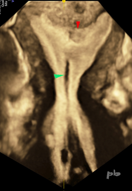
9 – Utérus à fond arqué
(U1c selon la classification de l’ESHRE)
En coupe transversale échographique, l’impression sur cette coupe est celle d’un utérus cloisonné avec 2 hémi cavités (★) et un fond utérin arrondi, sans incisure (►).
9 – Arcuate uterus (U1c according to ESHRE classification)
On transverse ultrasound, the impression on this slice is of a septate uterus with two hemi-cavities (★) and a rounded uterine fundus, without an indentation (►).

10 – Utérus à fond arqué
( U1c selon la classification de l’ESHRE)
(même patiente que 9)
Echographie endo-vaginale et reconstruction frontale après acquisition volumique.
Le fond utérin est arrondi, sans incisure (►).
La hauteur de la cloison est finalement très réduite (trait rouge). Elle correspond à la distance entre l’éperon en bas et l’horizontale tirée entre les 2 cornes (trait bleu). Elle est ≤ 50 % de l’épaisseur du myomètre, ce qui différencie cet utérus d’un vrai utérus cloisonné (U2).
10 – Arcuate uterus (U1c according to ESHRE classification)
(same patient as 9)
Transvaginal ultrasound with volumetric acquisition and frontal reconstruction. The rounded uterine fundus is confirmed, without indentation (►). The height of the septum is actually very minimal (red line). It corresponds to the distance between the spur at the base and the horizontal line drawn between the two horns (blue line). It is ≤ 50% of the myometrial thickness, which distinguishes this uterus from a true septate uterus (U2).
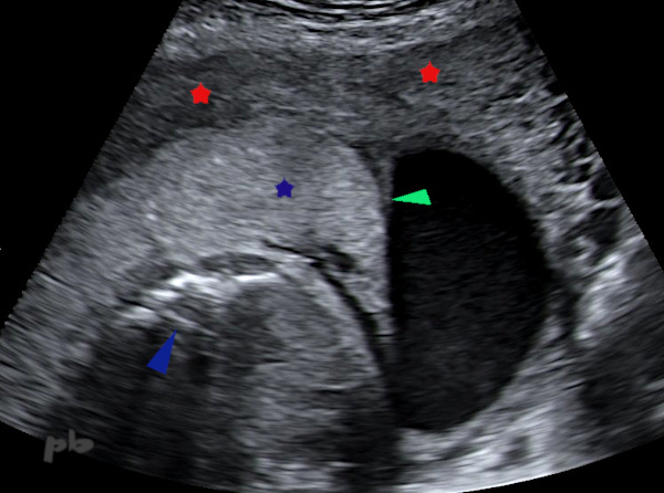
11 – Utérus cloisonné en cours de grossesse
Patiente à 28 SA.
Le placenta (★) vient s’insérer partiellement sur la cloison utérine (► qui était incomplète).
Myomètre (★).
Rachis fœtal (►).
11 – Septate uterus during pregnancy
Patient at 28 weeks of gestation.
The placenta (★) is partially inserted on the uterine septum (►, which was incomplete).
Myometrium (★).
Fetal spine (►).

12 – Utérus bicorne unicervical
(U3b C0 selon la classification de l’ESHRE)
Hystérographie (HSG).
Présence de 2 cavités (★) très divergentes. Le contour externe du fond utérin n’étant pas accessible en HSG (mais en échographie ou en IRM), on ne peut faire la différence entre un utérus bicorne (U3) et un utérus cloisonné (U2). Seul l’éperon du fond de la cavité est accessible (►), descendant jusqu’à l’orifice interne : division totale de la cavité (classement b).
L’HSG n’est donc plus réalisée dans ce contexte.
12 – Unicornuate bicornuate uterus (U3b C0 according to ESHRE classification) -Hysterosalpingography (HSG).
Presence of 2 highly divergent cavities (★). Since the outer contour of the uterine fundus is not accessible via HSG (though it is on ultrasound or MRI), it is impossible to distinguish between a bicornuate uterus (U3) and a septate uterus (U2). Only the spur of the fundal cavity is visible (►), extending down to the internal orifice : total division of the cavity (classification b).
HSG is therefore no longer performed in this context.
© Dr Philippe BASSNAGEL – 2022