
1 –Introduction vidéo
Séquence vidéo.
Un kyste dermoïde est souvent difficile à voir en échographie. Si vous ne l’avez pas vu, rendez-vous plus loin dans ce chapitre.
1 – Video Introduction
Video sequence.
A dermoid cyst is often difficult to visualize on ultrasound. If you haven’t seen it, refer to the later sections of this chapter.
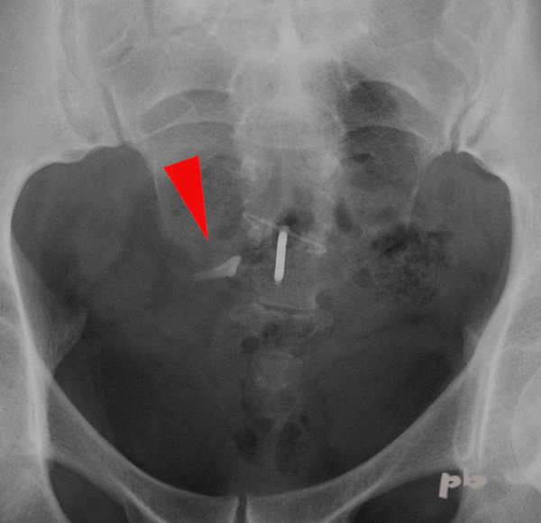
2 – Kyste dermoïde et ASP
Pendant très longtemps, l’ASP a été le seul moyen non invasif de diagnostiquer, souvent de manière fortuite, un kyste dermoïde.
Ici visualisation d’une dent (![]() ) à côté d’un DIU.
) à côté d’un DIU.
2 – Dermoid Cyst and Abdominal X-ray
For a very long time, abdominal X-ray was the only non-invasive way to diagnose—often incidentally—a dermoid cyst.
Here, visualization of a tooth ( ![]() ) next to an IUD (intrauterine device).
) next to an IUD (intrauterine device).
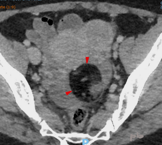
3 – Kyste dermoïde et TDM (scanner)
Comme l’ASP, le scanner qui utilise le rayonnement X, permet de visualiser les 2 tissus caractéristiques des kystes dermoïdes :
-les calcifications : elles sont dans la pratique rares
-la graisse, infiniment plus fréquente dans ces tumeurs, bien que non systématique. Si le kyste est suffisamment gros, il peut être visible sur un simple ASP par le contraste graisseux.
Kyste dermoïde typique (![]() ). Hypodensité en rapport avec le contenu graisseux.
). Hypodensité en rapport avec le contenu graisseux.
3 – Dermoid Cyst and CT Scan
Like a plain abdominal X-ray, the CT scan—which uses X-ray radiation—allows visualization of the two characteristic tissues of dermoid cysts:
*Calcifications: These are rare in practice.
*Fat, which is much more common in these tumors, though not always present. If the cyst is large enough, it may be visible on an abdominal X-ray due to the fat contrast.
Typical dermoid cyst ( ![]() ). Hypodensity corresponding to the fatty content.
). Hypodensity corresponding to the fatty content.
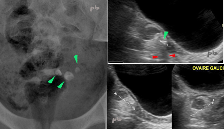
4 – Kyste dermoïde typique – ASP + échographie
Jeune fille de 12 ans.
ASP montrant 3 calcifications pelviennes gauches (![]() ). L’ASP n’est plus pratiqué actuellement (dossier ancien).
). L’ASP n’est plus pratiqué actuellement (dossier ancien).
Echographie par voie sus-pubienne : image ovarienne gauche (entre les croix) avec une composante hyperéchogène et une 2ème hypoéchogène. Elles correspondent à du sébum et à des phanères.
Une calcification (![]() ) avec cône d’ombre (
) avec cône d’ombre (![]() ).
).
Vessie pleine en noir.
4 – Typical Dermoid Cyst – Plain Abdominal X-ray + Ultrasound
12-year-old girl. Plain abdominal X-ray showing three left pelvic calcifications ( ![]() ). Note: It is no longer currently performed (old case file). Suprapubic ultrasound: Left ovarian image (between the crosses) with a hyper-echoic component and a second hypo-echoic component. These correspond to sebum and hair. A calcification (
). Note: It is no longer currently performed (old case file). Suprapubic ultrasound: Left ovarian image (between the crosses) with a hyper-echoic component and a second hypo-echoic component. These correspond to sebum and hair. A calcification ( ![]() ) with an acoustic shadow (
) with an acoustic shadow ( ![]() ).
).
Full bladder appears black.
© Dr Philippe BASSNAGEL – 2022